|
|
Pulmonary Alveolar Microlithiasis
General Considerations
- Very rare, chronic disease characterized by widespread calcifications called "calcispherites" within the alveoli
- Etiology is unknown but it has a familial incidence with an autosomal recessive inheritance pattern
- No known calcium metabolism disorder
- Microliths range from 0.01 to 3 mm in size and are composed of calcium phosphate
Clinical Findings
- May remain asymptomatic until 3rd-4th decade
- First symptom is usually dyspnea on exertion
- Restrictive lung disease pattern
- Eventually, there may be cyanosis and clubbing
Imaging Findings
- Diffuse, discrete, very small, symmetric micronodular calcifications (sand-storm appearance)
- Predominantly in mid- to lower-lung zones with sparing of apices
- Heart borders and diaphragm are obliterated from view
- Denser subpleural layer
- "Black pleural line"
- Increased lucency between dense lung parenchyma and ribs
- Small apical bullae
- High-resolution computed tomography is the study of choice
- Diffuse ground-glass opacities
- May be more prominent centrally than peripherally
- Densely calcified, diffusely spread calcified nodules
- May show pleural calcification as well
Differential Diagnosis
- Same "crazy-paving" described with alveolar proteinosis may be seen in microlithiasis
- Silicosis
- Metastatic calcification in renal disease
Treatment
Complications
- Respiratory insufficiency
- Cyanosis
- Clubbing
- Pulmonary hypertension
Prognosis
- Microliths may continue to form or disease may arrest


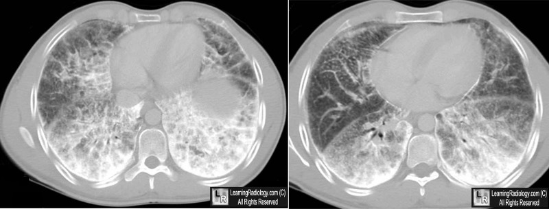
Pulmonary Alveolar Microlithiasis. Axial CT scans of the lower chest demonstrate extremely dense
calcific material at both lung bases (red arrows) which is also producing pleural calcification (black arrows). The
disease is seen to be composed of innumerable smaller calcifications (white circle).
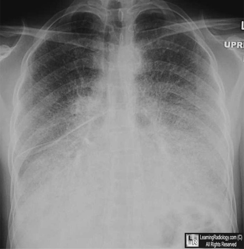
The black arrow on the conventional chest radiograph points to the lower lobe predominance.
The
blue arrow points to calcification of the fissures.
For more information, click on the link if you see this icon 
For these same photos without the annotations, click here and here
Pulmonary alveolar microlithiasis presenting with crazy-paving pattern on high resolution CT. British Journal of Radiology (2004) 77, 974-976. E L Gasparetto, MD; P Tazoniero, MD; D L Escuissato, MD; E Marchiori, MD; R L Frare e Silva, MD and D Sakamoto, MD.
Case report - pulmonary alveolar microlithiasis. MS Shah, KI Nanavati, N Airon, RR Shah, BD Joshi. CHEST: 2003 | Volume : 13 | Issue : 3 | Page : 277-279
|
|
|
