|
|
Monad's Sign and Air Crescent Sign
General Considerations
- Sickle-shaped lucency, partially surrounding a soft tissue mass in a pulmonary cavity seen on conventional radiography and CT
- Terminology can be confusing as a similar sign is described for both invasive aspergillosis and secondary, non-invasive, saprophytic aspergillosis
- Invasive aspergillosis typically occurs in immunocompromised patients
- It frequently begins with a solid-appearing peripheral soft tissue opacity that, through infarction and retraction, produces a cavity with a density within it
- The density is necrotic lung tissue, not the fungus ball in non-invasive aspergillosis
- Sputum cultures are not often positive (10%) so diagnosis may require bronchoscopy or percutaneous aspiration
- Saprophytic aspergillosis (aspergilloma, mycetoma, fungus ball) occurs in a pre-existing cavity
- It is more common than the invasive form
- Usually occurs in a cavity from TB or sarcoid
- The "ball" is composed of hyphae, mucous and cellular debris
- Hemoptysis is a common symptom
Imaging Findings
- In secondary, non-invasive (saprophytic) aspergillosis
- The fungus ball is usually freely movable and will move to the dependent surface on decubitus radiographs or supine and prone CT studies
- Rarely, the fungus ball may calcify
- Invasive aspergillosis
- On CT, there may be ground glass densities in the area around the nodular density called the "halo sign" and indicative of hemorrhage
Differential Diagnosis
- Cavitating neoplasm
- TB
- Nocardiosis
 
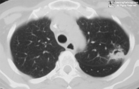
Monad's sign. The axial CT scan of the chest, on the left, shows a nodule (black arrow) within an air-filled cavity with a crescent of air at the superior surface of the cavity (white arrow). In the close-up view of the left upper lobe from a chest radiograph of another person, on the right, there is again seen a thin-walled cavity (white arrow) containing a soft-tissue mass (black arrow). Both of these cavities were from old tuberculosis.
For more information, click on the link if you see this icon 
For this same photo without the annotations, click here or here
Abramson, S. January 2001 Radiology, 218, 230-232
Saprophytic aspergillosis Eurorad case 6672
|
|
|
