|
Histoplasmosis
· Caused by Histoplasma Capsulatum, a soil fungus found especially in the Ohio River Valley (Ohio, Mississippi)
o Infected soil can be carried by birds, although birds themselves are not infected
· Similar in course and imaging appearance to tuberculosis
· Histoplasmosis is the most common cause of fibrosing mediastinitis
· Common cause of diffuse punctate splenic calcifications
o Spleen is more frequent site of calcifications than liver
o DDX: Brucellosis, TB, Candidiasis in immunocompromised patients
§ Number of calcifications: more than 6, more likely histoplasmosis
§ Size of calcifications: larger the size, the more likely histoplasmosis
§ Appearance of calcifications: in Brucellosis, calcifications may be 1-3 cm in size (larger than in Histo or TB), and have a calcified rim surrounding a lucent center
· There also may be suppurative lesions of the spleen present at the same time as the calcifications, unusual for Histo or TB
· Diffuse uniform calcification of the spleen is most often seen in sickle cell disease
o Thorotrast may mimic diffuse calcification but is of a higher density and is usually present in abdominal lymph nodes as well
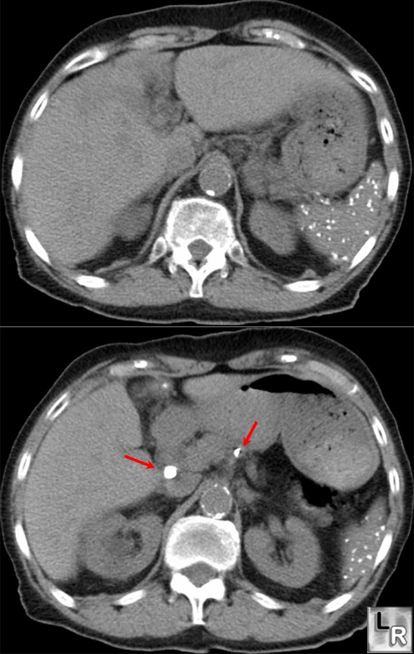
Histoplasmosis. There are numerous, punctate calcifications in the spleen and calcification in
two mesenteric lymph nodes. TB usually produces fewer splenic calcified granulomas than histoplasmosis.
For a photo of the same image without arrows, click on this link
· Clinical findings
o Portal of entry is the respiratory tract
o High male to female ratio (4:1)
o Most infections are asymptomatic
§ Fewer than 5% of those infected will develop symptoms
§ Primary risk factors for development of symptoms are age and immunosuppression
o Tissues react to infection by forming caseating and/or non-caseating granulomas which may calcify
Primary histoplasmosis
· Most infections are subclinical
o Most acute symptomatic cases are mistaken for a flu-like illness
· The lungs are the most common organ affected
· Chest radiographs are most often normal but may show adenopathy with a focal area of consolidation
· Healing can occur with formation of a histoplasmoma
o Histoplasmoma=target calcification=bull’s-eye calcification in center of nodule (Pathognomonic)
Chronic histoplasmosis
· Chronic pulmonary histoplasmosis is more likely to occur in those with underlying lung disease like COPD
· Imaging findings
o Migratory bronchopneumonia
o Hilar and mediastinal adenopathy
o Multiple nodules which heal with numerous calcified granulomas
§ TB granulomas are usually 1 or 2 in number
o Upper lobe cavitation, healing with fibrosis (very similar to TB)
Progressive Disseminated Histoplasmosis
· Affects multiple organs
· Infants, elderly and immunosuppressed are predisposed
o CD4 count is usually below 150-200/mm
· Most cases believed to arise from re-activation
· Can be rapidly fatal
· Imaging
o Consolidation
§ Diffuse and/or miliary pattern
o Cavitation
§ Upper lobe, most often in men with COPD
o Adenopathy
§ Mediastinal or hilar
o Hepatosplenomegaly
o Lytic lesions in the bones
o Broncholithiasis (most common cause)
o Fibrosing mediastinitis
o Multiple splenic calcifications (unusual for TB)
o Epididymitis and prostatitis
Non-pulmonary histoplasmosis
· Pericarditis
· Arthralgias
· Sarcoid-like syndrome in which the histopathologic findings are similar to sarcoid and, like sarcoid, ACE levels are high
|
