|
|
Pseudocoarctation of the Aorta
General Considerations
- Congenital and relatively rare
- Elongated and redundant descending aorta with kinking or buckling of the aorta distal to the origin of the left subclavian artery
- No obstruction
- Pressure gradient of less than 30 mmHg
- No rib notching
- May be caused by an abnormally long descending aorta
Clinical Findings
Imaging Findings
- Junction of arch and descending aorta just past ductus
- May demonstrate "Figure 3 sign"
- Since there is no collateral flow needed, there is no rib notching
Differential Diagnosis
- True adult coarctation of the aorta
- Pressure differences and rib notching
- Cervical aortic arch
- Usually descends on the side opposite the arch
Treatment
- No specific treatment for the pseudocoarctation
Complications
- Associated with bicuspid aortic valve, aortic stenosis and insufficiency, PDA, VSD and corrected transposition

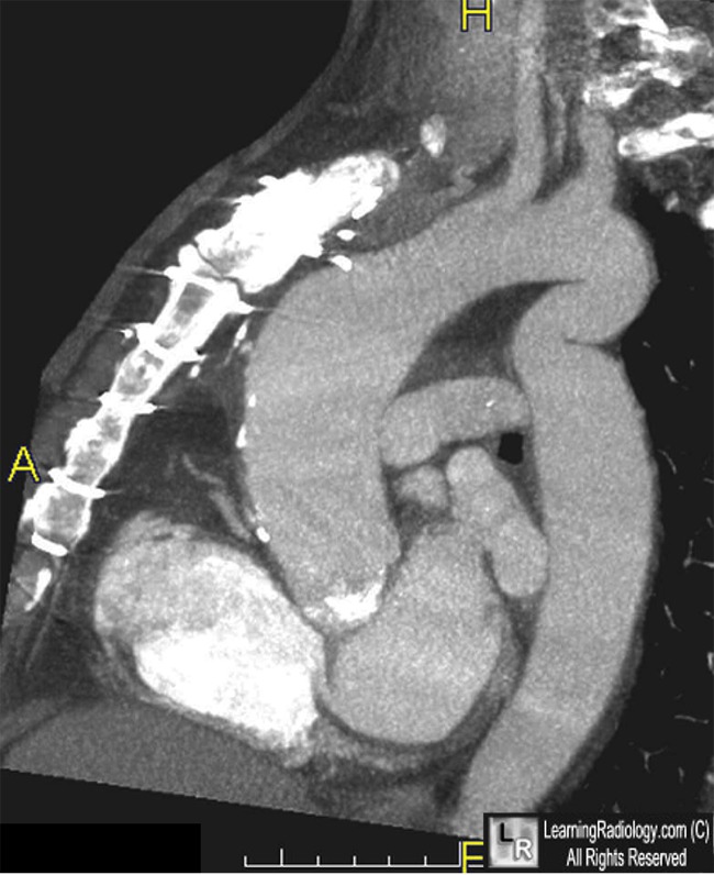
Sagittal CT of aorta

Pseudocoarctation. Sagittal contrast-enhanced CT (above) shows kinking of aorta (white arrow)
that produces a picture similar to coarctation without the significant pressure gradient seen in a true coarctation.
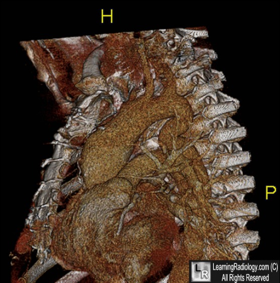
A surface rendered 3D image below shows the same abnormality. AA=ascending aorta; PA=pulmonary artery.
For these same photos without the arrows, click here and here
For more information, click on the link if you see this icon 
Pseudocoarctation of the aorta. Bluemke DA. Cardiology Journal. Vol. 14, No. 2, pp. 205.206, 2007.
|
|
|
