|
|
Ductus Diverticulum
Ductus Bump
General Considerations
- Occurs on the anterior and medial surface of the aortic isthmus at the site of the ductus arteriosus
- Found in about 9% of adults on aortography
- More commonly found in children
Clinical Findings
Imaging Findings
- Best imaged with contrast-enhanced Ct with 3D reconstruction or MRI
- On CT, best visualized on sagittal oblique reconstructed images
- Diverticulum has smooth and uninterrupted margins
- Obtuse angles formed by the ductus diverticulum and the aortic wall
- More atypically, a ductus diverticulum may have a steeper slope on its cephalad margin and a gentle slope on its inferior margin
Differential Diagnosis
- Pseudoaneurysm (forms acute angle with aorta)
- On aortography, will demonstrate an intimal flap and/or
- Delayed clearance of contrast
- Penetrating ulcer
- Aortic dissection
Treatment
- Usually unnecessary unless diverticulum is greater than 3 cm
Prognosis
- No fatal sequelae usually
- Rare formation of larger aneurysm

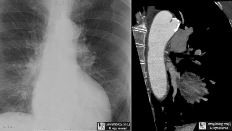
Ductus Diverticulum. The chest radiograph demonstrates a rim-calcified lesion adjacent to the aortic knob (white arrow). CT reconstruction demonstrates a projection from the anterior wall of the aorta at the level of the ligamentum arteriosus (blue arrow) The diverticulum is atypical in that it has a steeper angle cephalad.
For this same photo without the arrows, click here
For more information, click on the link if you see this icon 
|
|
|
