|
|
Congenital Defect of The Pericardium
General Considerations
- Rare absence of a part or all of the pericardium
- Due to failure of pericardial development secondary to premature atrophy of the left duct of Cuvier (cardinal vein) which then fails to nourish the left pleuropericardial membrane
- Male:female ratio of 3:1
- May be detected at any age but most commonly in low 20’s
Location:
- Foraminal defect on left side 35%
- Complete absence of left side 35% —levoposition of heart
- Diaphragmatic surface 17%
- Total bilateral absence 9%
- Right sided 4%
Clinical findings
- Mostly asymptomatic
- May have:
- Tachycardia
- Palpitations
- Right bundle block
- Positional discomfort lying on left side
- Chest pain
Imaging Findings
- Focal bulge in area of main pulmonary artery or left atrium in partial defects
- In complete form, heart rotates up and to the left
- Lung may interpose between heart and left hemidiaphragm
- Increased distance between sternum and heart 2° absence of sternopericardial ligament
- Lung may interpose between aorta and main pulmonary artery on axial CT scans
- Levoposition of heart
- Pneumopericardium may occur following pneumothorax
Treatment
- Asymptomatic, complete absence of the pericardium and very small defects present no danger to the patient and require no intervention
- Most authors advise leaving incidental defects untreated
- Symptomatic foraminal defect may require surgery because of herniation and strangulation of left atrial appendage or herniation of LA/LV
- Surgery can be enlargement of the defect to prevent strangulation or closure of the defect
Complications and associations
- Associated congenital anomalies occur in about 30 per cent of the reported cases
- Bronchogenic cysts
- ASD, VSD, PDA, Tetralogy of Fallot, Mitral stenosis
- Diaphragmatic hernia
- Pulmonary sequestration

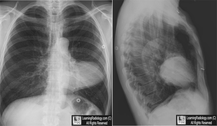
Congenital Defect in the Pericardium. Frontal and lateral chest radiographs demonstrate an unusually-shaped (yellow arrow) levopositioned (green arrow) heart. The heart is displaced upward from the left hemidiaphragm (white arrow) and there is a clear space between the sternum and the heart (blue arrow) on the lateral image.
For more information, click on the link if you see this icon 
For these same photos without the annotations, click here
Congenital Defects of the Pericardium. Nigel E. Drury, Ravi J. De Silva, Roger M. O. Hall and Stephen R. Large. Ann Thorac Surg 2007;83:1552–3
|
|
|
