|
Synovial Chondromatosis
Synovial Osteochondromatosis
· Occurs in two forms: primary and secondary
· Primary synovial chondromatosis is an uncommon disease in which there is metaplasia of the synovial lining of joints, bursae or tendons into cartilaginous nodules
o The nodules may detach and become “loose bodies” in the joint
o As the loose bodies receive their nourishment from the synovial fluid, they may continue to grow even though “floating” in the joint
· Usually monarticular
· Much more common in men than women
· Usually in 3rd-5th decade
· Knee is most frequent site of involvement, followed by
o Elbow
o Hip
o Shoulder
o Ankle
o Wrist
· Clinical findings
o Pain
§ Usually chronic and progressive
o Swelling
o Limitation of movement (lock knee)
o Joint effusion
§ May be hemorrhagic
o Occasionally it can present as a slow-growing soft tissue mass
· Imaging findings
o Depend in large part on the degree of calcification of the cartilaginous bodies
§ 1/3 of chondromas show no radio-opacity
o An important negative fining in primary form is that the adjacent joint is usually normal
o The nodules are usually uniform in size ranging from a few mm to several cm
o Small
o Multiple
o There may be pressure erosion of adjacent bone in joints
o Rarely, widening of joint space from accumulation of loose bodies
o Usually no osteoporosis
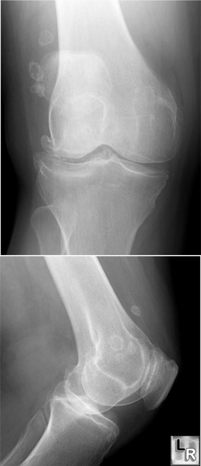
Secondary form of Synovial Osteochondromatosis. There are several osteocartilaginous
nodules seen around the knee joint space. They are relatively few in number and associated with
osteoarthritis of the knee. This is the appearance of secondary synovial osteochondromatosis.
For a large photo of the same image, click on this link
Primary form of Synovial Osteochondromatosis. There are numerous osteocartilaginous
nodules seen around the knee joint space. They are relatively uniform in size and are not associated with
osteoarthritis of the knee. This is the appearance of primary synovial osteochondromatosis.
· CT findings
o Intra-articular soft-tissue mass of near water attenuation containing multiple small calcifications
· MRI findings
o Lobulated intra-articular mass isointense to muscle on T1WI and hyperintense to muscle on T2WI containing multiple foci of low signal intensity
· Rarely, they may degenerate into a chondrosarcoma
o Frequently marked by the onset of pain and an associated soft tissue mass
· Treatment
o Removal of loose bodies
o May recur
· Secondary osteochondromatosis
o Caused by and associated with osteoarthritis
o Most common in knee and hip
o Loose bodies are fewer in number and less uniform in size than in primary form
· Synovial chondrosarcoma
o May resemble synovial osteochondromatosis
o Clues may include destruction of bone and an associated soft tissue mass
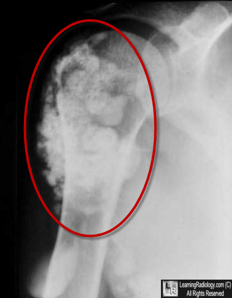
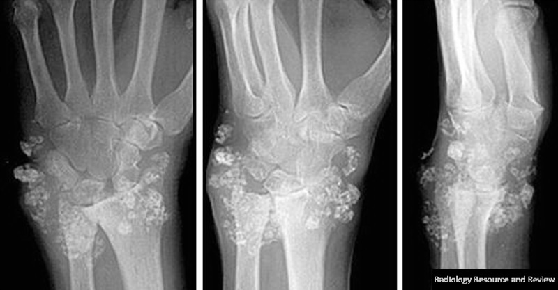
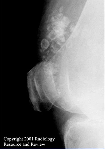
Top: Multiple nodular calcifications surrounding the right shoulder in synovial osteochondromatosis. Bottom: Multiple nodular calcifications surrounding the wrist in the same disease.
|
