|
|
Stress Fracture of the Foot
General considerations
- Fractures produced as a result of repetitive stress on bone
- General risk factors
- New, different or rigorous repetitive activity
- Females
- Increasing age
- Caucasians
- Low bone mineral density
- Low calcium intake
- In the foot, these are most common in the shafts of the 2nd (most) and 3rd metatarsals
- Marching, stomping on ground, prolonged standing, ballet, postoperative bunionectomy increase risk
Clinical findings
- Pain usually starts more diffusely and then localizes to site of fracture
- Swelling
- Redness
- Localized tenderness
Imaging findings
- Conventional Radiography
- 15% sensitive in early fractures, increasing to 50% on follow-up
- Sclerotic band (due to trabecular compression and callus formation) usually perpendicular to cortex
- Intracortical radiolucent striations (early)
- Solid thick lamellar periosteal new bone formation
- Endosteal thickening (later)
- Follow-up radiography after 2-3 weeks of conservative therapy may reveal fracture not seen earlier
- Nuclear medicine
- “Gold standard" = almost 100% sensitive
- Abnormal uptake within 6-72 hours of injury (prior to radiographic abnormality)
- "Stress reaction" is a focus of subtly increased uptake
- Focal fusiform area of intense cortical uptake
- Abnormal uptake persists for months
- MRI
- Very sensitive modality
- Fat saturation technique most sensitive to detect increase in water content of medullary edema / hemorrhage
- Diminished marrow signal intensity on T1WI
- Increased marrow signal intensity on T2WI
Prognosis
- Usually heal without complication when involving 1st through 4th metatarsals
Complications
- Nonunion
- Higher (up to 50%) in 5th metatarsal fracture

Stress fracture of the 3rd metatarsal. There is periosteal reaction (white circle) at the site of a stress fracture of the shaft of the 3rd metatarsal. The fracture itself is not visible but the periosteal reaction (callus formation) is.
For this same photo without the annotations, click here
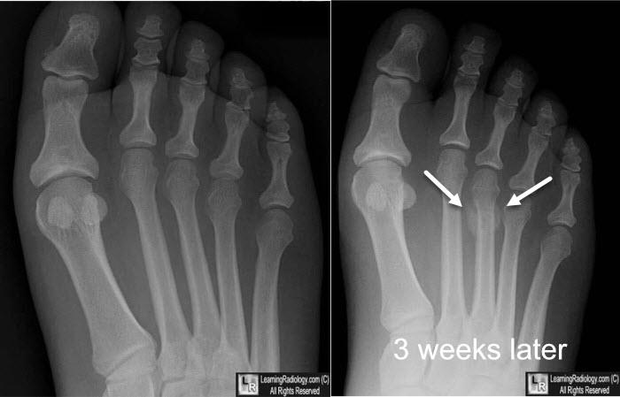
Stress fracture of the 3rd metatarsal. At the time of injury (image on left) there is no visible abnormality. Three weeks later there is periosteal reaction (white arrows) at the site of a stress fracture of the shaft of the 3rd metatarsal. The fracture itself is not visible but the periosteal reaction (callus formation) is.
|
|
|
