|
|
Salter-Harris Fractures
Salter III
- General Considerations
- The epiphyseal plate (physis or growth plate) is the weakest part of the bone to shearing injuries
- The Salter-Harris classification is a means of categorizing epiphyseal plate fractures and provides clues to their prognosis
- All such these fractures, by definition, involve or extend through the epiphyseal plate so that all such fractures occur in children before the epiphyseal plate closes
- Salter-Harris I Fractures

- Occurs through the hypertrophic zone of the epiphyseal plate
- Only the epiphyseal plate is fractured
- Rarely produces complications
- May be difficult to diagnose unless there is visible displacement of the epiphysis on the metaphysis
- Slipped capital femoral epiphysis (SCFE) is an example of a Salter-Harris I fracture
- Salter-Harris II Fractures
- Most common Salter-Harris fracture -85%
- Involves both the epiphyseal plate and the metaphysis
- Small corner of metaphysis that is usually fractured produces the “corner sign”
- Rarely produces complications
- Salter-Harris III Fractures
- Involves the epiphyseal plate and the epiphysis itself
- Since the epiphysis is involved, damage to the articular cartilage can occur
- Growth disturbance is uncommon
- A Tillaux fracture of the ankle is a Salter-Harris III fracture
- Salter-Harris IV Fractures
- Involves the epiphyseal plate, metaphysis and epiphysis
- Since it, too, involves the epiphysis, the articular cartilage can be damaged
- Since these fractures involve the growing layer of cartilage, growth disturbance can result
- Salter-Harris V Fractures
- Rare
- Compression or crushing injury of epiphyseal plate
- Initial diagnosis may be difficult and not made until complication of growth disturbance at epiphyseal plate occurs resulting in angular deformities
- Associated with growth disturbance
- These injuries have the worst prognosis of the Salter-Harris fractures
Structures involved in Salter-Harris fractures
|
Type |
Involves epiphyseal plate |
Fracture of metaphysis |
Fracture of epiphysis itself |
I |
Yes |
|
|
II |
Yes |
Yes |
|
III |
Yes |
|
Yes |
IV |
Yes |
Yes |
Yes |
V |
Yes |
|
|
- Clinical Findings
- Point tenderness
- Pain
- Swelling
- Limitation of motion
- Imaging Findings
- Soft tissue swelling
- Depending on the type of fracture, some displacement of the epiphysis or corner sign (Thurston-Holland fragment)
- Conventional radiography remains study of first choice
- CT with multiplanar reconstruction has been used in problem cases
- Ultrasound can be helpful in infants whose cartilage has not yet ossified
- MRI in problem cases
- Complications
- Complications are rare
- In general, the higher the number, the more likely the complication so that Salter-Harris types Iv and V have the highest associated complications
- Greater risk for complication comes with fracture of distal tibia followed by distal femur
- Primary complication is growth plate disturbance
- Early closure
- Closure of only a portion of the plate resulting in angular deformity

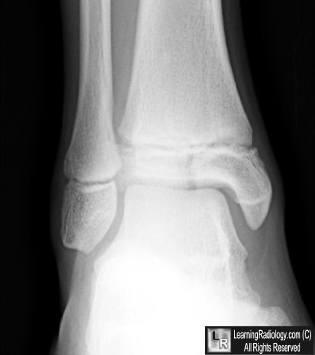
Salter-Harris III Epiphyseal Fracture. There is a longitudinal lucency (blue arrow) in the
epiphysis that represents a fracture. All Salter-Harris fractures, by definition,
involve the epiphyseal plate, even though those fractures may not be visible.
For this same photo without the arrows, click here
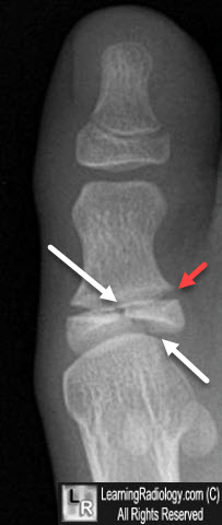
Salter-Harris III Epiphyseal Fracture. There is a longitudinal lucency (whie arrows) in the
epiphysis of the great toe that represents a fracture. All Salter-Harris fractures, by definition, involve the
epiphyseal plate (red arrow), which is wider than normal in this case.
For more information, click on the link if you see this icon 
|
|
|
