|
|
Acute Osteomyelitis
- Age
- Usually affects children
- Septic arthritis more common in adults;
osteomyelitis in children
Hallmark characteristics
- Destruction of bone
- Periosteal new bone formation
- Organisms
- Newborns
- S. aureus
- Group B streptococcus
- E. coli
- Children
- Adults
- S. aureus (most common)
- Enteric species
- Streptococcus
- Drug addicts
- Pseudomonas (most common)
- Klebsiella
- Sickle cell disease
- Pathogenesis
- Hematogenous spread
- Direct implantation from a traumatic /
iatrogenic source
- Extension from adjacent soft-tissue infection
- Location
- Lower extremity (most common)
- Over pressure points in diabetic foot
- Vertebrae
- Lumbar > thoracic > cervical
- Radial styloid
- Sacroiliac joint
ACUTE NEONATAL OSTEOMYELITIS
- Age
- Little or no systemic disturbance
- Multicentric involvement more common
- Bone scan falsely negative / equivocal in 70%
ACUTE OSTEOMYELITIS IN INFANCY
- Age
- Pathomechanism
- Spread to epiphysis through blood vessels
- Marked soft-tissue component
- Subperiosteal abscess with extensive periosteal
new bone formation
- Complications
- Frequent joint involvement
- Prognosis
ACUTE OSTEOMYELITIS IN CHILDHOOD
- Age
- Pathomechanism
- Trans-physeal vessels closed
- Primary focus of infection is in metaphysis
- Findings
- Sequestration frequent
- Periosteal elevation
- Small single / multiple osteolytic areas in
metaphysis
- Extensive periosteal reaction parallel to
shaft (after 3-6 weeks)
- Shortening of bone with destruction of
epiphyseal cartilage
- Growth stimulation by hyperemia and premature
maturation of adjacent epiphysis
ACUTE OSTEOMYELITIS IN ADULTHOOD
- Delicate periosteal new bone
- Joint involvement common
- X-ray findings
- Initial radiographs often normal for as long
as 7-10 days
- Localized soft-tissue swelling adjacent to
metaphysis with obliteration of usual fat planes (after 3-10 days)
- Area of bone destruction (lags 7-14 days
behind pathologic changes)
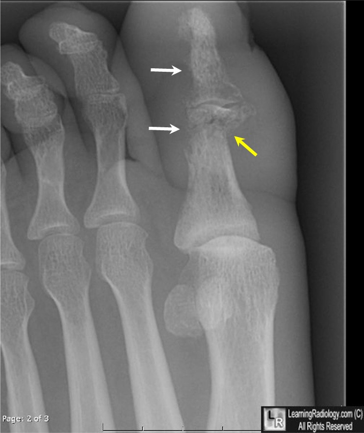
Osteomyeltis. Bone
destruction of proximal and terminal phalanges of great toe with destruction of the cortex
characteristic of osteomyelitis (white arrows). There is also a pathologic fracture through the proximal phalanx (yellow arrow)
- Involucrum = cloak of laminated /spiculated periosteal reaction (develops after 20 days)
- Sequestrum = detached necrotic cortical bone
(develops after 30 days)
- Cloaca formation =
space in which dead bone resides
- MR findings
- Bone marrow hypointense on T1WI + hyperintense on T2WI (= water-rich inflammatory tissue)
- DDx
- Neuropathic osteoarthropathy
- Aseptic arthritis
- Acute fracture
- Recent surgery
- Ewing’s sarcoma
- Findings
- Focal / linear cortical involvement
hyperintense on T2WI
- Hyperintense halo surrounding cortex on T2WI =
subperiosteal infection
- Hyperintense line on T2WI extending from bone
to skin surface and enhancement of borders (= sinus tract)
- Nuclear Medicine (accuracy approx. 90%):
- Ga-67 scans
- 100% sensitivity
- Increased uptake 1 day earlier than for
Tc-99m MDP
- Gallium helpful for chronic osteomyelitis
- Static Tc-99m diphosphonate
- 83% sensitivity
- 5-60% false-negative rate in neonates and
children
- Complications of osteomyelitis
- Abscess in soft-tissue
- Fistula or sinus formation
- Pathologic fracture
- Extension into joint producing septic
arthritis
- Growth disturbance due to epiphyseal
involvement
- Severe deformity with delayed treatment
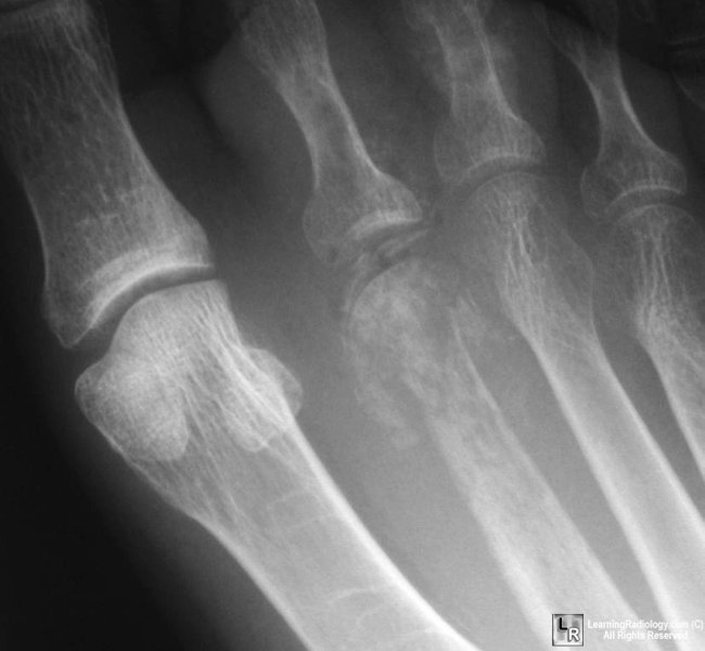
Osteomyeltis. Bone
destruction of head of 2nd metatarsal
with periosteal new bone formation characteristic of osteomyelitis
|
|
|
