|
|
Legg-Calve-Perthes Disease
Idiopathic Osteonecrosis of the Capital Femoral Epiphysis
General Considerations
- Idiopathic avascular necrosis of the femoral head
- Peak incidence is five years with a range of 2-14 years
- Occurs in males greater than females, more often in Caucasians
- Bilateral 10-20% of time. Most often one after the other rather than simultaneously
- Bilateral involvement in females is very rare
- Predispositions
- Males
- Low birth weight
- Lower socio-economic groups
- Presence of inguinal hernia
- GU tract anomalies
Clinical Findings
- Intermittent limp, especially after exertion
- Pain in thigh
- Limited range of motion
- Hip pain which may be referred to the knee
Imaging Findings
- Conventional radiography is the primary modality of diagnosis and upon which staging is based (97% sensitivity and 78% specificity)
- Small capital femoral epiphysis (early)
- Sclerosis of head
- Widening of the joint space from joint fluid ligamentous laxity
- Destruction of the articular cortex similar to septic arthritis
- Crescent sign-subarticular lucency from subchondral fracture (late sign)
- Fragmentation of femoral head
- Bone scan may be used early in the disease as may be MRI
- Low signal replaces normal high signal in femoral epiphysis on T1 and T2
- Intra-articular effusion
- Femoral head deformity
- On Bone scan
- Photopenic area in proximal femoral epiphysis because of interruption of blood supply to epiphysis
- CT allows for early diagnosis as well
- Collapse of head
- Curvilinear sclerotic zone
- Areas of decreased attenuation in head
- Intraosseous cysts
Differential Diagnosis
Treatment
- More than 50% do not require treatment
- Initial therapy includes minimal weight-bearing
- Bracing or surgery to maintain abduction and rotation of hip
- With revascularization there is gradual reformation of femoral head
Complications
- Early osteoarthritis
- Limp
Prognosis
- Better in those in whom the disease develops before 10 years of age and best in those < 5 years


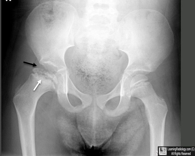
Legg-Calve-Perthes Disease. AP and frog-lateral views of both hips show a right capital
femoral epiphysis which is smaller in size (black arrows) than the left (white arrow), an early sign of this disease.
For these same photos without the arrows, click here and here
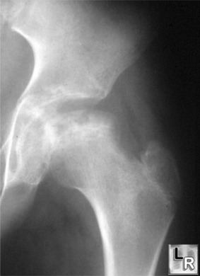
Frontal radiograph of left hip in a 3
year-old shows flattening,
sclerosis and cystic lucencies with preservation of the hip joint space
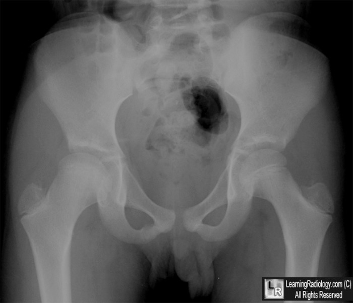
Legg-Calve-Perthes Disease. AP view of both hips show a right capital
femoral epiphysis which is fragmented and collapsed (white arrow) while the joint space, as is characteristic at this stage, remains intact (black arrow).
For more information, click on the link if you see this icon 
Legg-Calve-Perthes Disease Imaging. eMedicine. AN Khan, DM Seriki, CE Hutchinson, S MacDonald.
|
|
|
