|
|
Ivory Vertebra
Vertebra Nigra
General Considerations
- Usually homogeneous, increased density of a vertebral body
- When conventional radiographs were displayed with black and white inverted (Britain), it was also called "vertebra nigra"
- Most often due to metastases in adults
- Metastases are most common malignant bone tumors
- Most involve axial skeleton
- Spread hematogenously
- Most frequently occur where red bone marrow is found
- Mets to spine frequently destroy posterior vertebral body including pedicle first=”pedicle-sign"
- 90% of skeletal mets are multiple
- Primary carcinomas that frequently metastasize to bone
- The next four lesions comprise 80% of all metastases to bone
- Breast (70% of bone mets in women)
- Lung
- Prostate (60% of all bone mets in men)
- Kidney
- Also
- Thyroid
- Stomach and intestines
Clinical Findings
- Most lesions are asymptomatic
- When symptomatic, pain is major symptom
Imaging Findings
- Metastases that are usually purely blastic (see chart below)
- Prostate
- Medulloblastoma
- Bronchial carcinoid
Causes of an Ivory Vertebra
|
Metastatic Disease from Prostate, Breast, Colon |
Lymphoma |
Medulloblastoma |
Paget Disease (“picture framing”) |
Hemangioma |
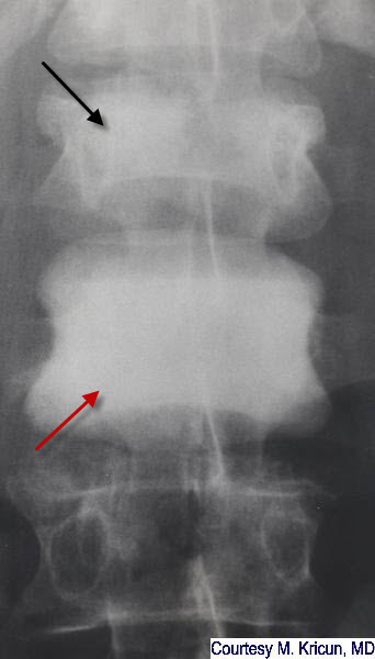
Ivory Vertebra. Markedly dense lumbar vertebral body (red arrow) that maintains its normal height but is increased in opacity. Another, compressed vertebral body with increased density is seen above it (black arrow). The patient had metastatic disease from a known colon cancer.
Orthopedic radiology: A Practical Approach, Greenspan, Ada; Lippincott, 2000
Diagnosis of Bone and Joint Disorders, Resnick, Donald, W. B. Saunders
Musculoskeletal Imaging: The Requisites, Manaster, BJ et al; Mosby, 2002
|
|
|
