|
|
Freiberg Infraction
Avascular Necrosis, Osteonecrosis, Osteochondrosis
General considerations
- Named “infraction” because it was originally thought secondary to trauma
- Exact cause remains uncertain but thought to be one of the osteochondroses in adolescents
- Osteochondroses are diseases that usually affect the epiphyses of growing bones resulting in necrosis most likely on a vascular basis, although the exact mechanism is not known
- In others, Freiberg's may be due to a combination of trauma, and vascular insults
- Relatively uncommon
- Painful collapse of the head of the 2nd metatarsal
- May affect 3rd metatarsal head as well
- Women to men by 5:1
- Possibly because of shoes, i.e. stresses placed on toe by high-heeled shoes
- Length of second metatarsal thought to be a factor by some
- Usually adolescents
- Almost always unilateral
Clinical findings
- Local pain, activity-related
- Tenderness
- Stiffness and limp
Imaging findings
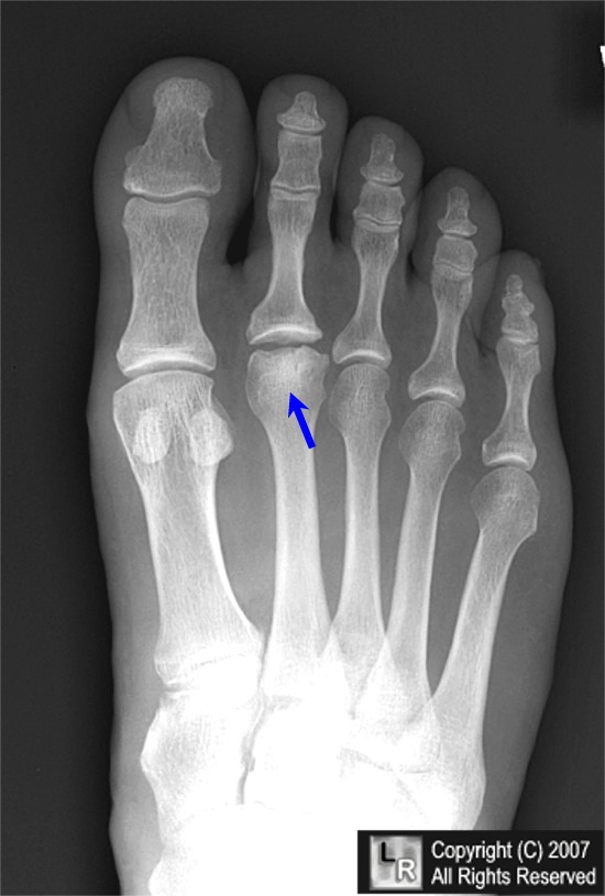
- Early signs are sclerosis of 2nd MT head and widening of joint space
- Later there is fragmentation and collapse
- End result is flattening of head
- May produce “loose body”
Treatment
- Medical
- Immobilization and avoidance of weight-bearing to rest the joint
- Surgical
- Various osteotomies, bone grafts, excision of the head, joint replacement have each been used alone or in combinations
Complications
- Premature closure of growth plate
- Loose bodies
- Secondary osteoarthritis
Other osteochondroses
- Kohler's disease of the tarsal navicular
- Panner's disease of the capitellum
- Sever's disease of the calcaneal apophysis
- Legg-Calve-Perthes disease of the capital femoral epiphysis

- Kienbock’s disease of the lunate

- Scheuermann’s disease of the spine

Freiberg's Infraction. Frontal radiograph of the foot demonstrates flattening and sclerosis of the head of the
2nd metatarsal classical for this disease. The base of the proximal phalanx is intact and the joint space appears widened.
For additional information about this disease, click on this icon above.
For this same photo without the arrows, click here
EMedicine Freiberg Infraction Boyer, M, DeOrio, JK
|
|
|