|
|
Fibrous Dysplasia
-
Etiology
unknown
-
Most common 3-15
years
-
Fundamental
abnormality is replacement of medullary bone by fibrous tissue
-
Clinically
-
Most commonly
involved bones are pelvis, femora
-
In widespread
disease, the skull and jaw are almost always involved
-
Imaging
-
Prone to
fracture
-
Growth of
lesions usually stops when epiphyses close
-
DDX:
-
Brown tumor
-
Unicameral
bone cyst
-
Albright’s
Syndrome
-
Polyostotic
form is usually bilateral
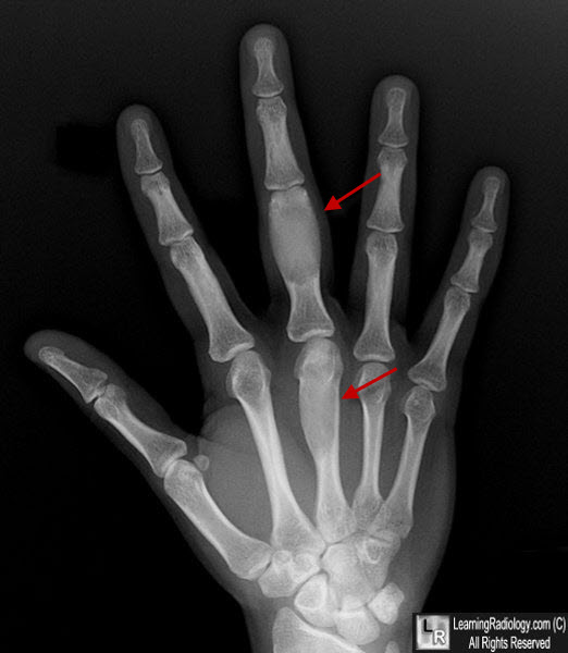
Fibrous Dysplasia. There are expansile, geographic, lytic lesions in the shafts of the 3rd metacarpal and proximal phalanx of the middle finger (red arrows). They have a characteristic "ground-glass" appearance to their internal matrices. Enchondromas can have a similar appearance though their internal matrix would be more coarse, resembling rings and arcs.
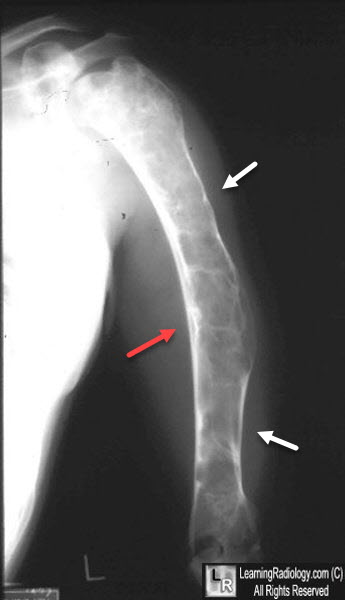
Fibrous Dysplasia. There are expansile, geographic, lytic lesions in the shaft of the humerus (white arrows) with endosteal scalloping and cortical thinning. The arm is bowed (red arrow). The matrix has a ground-glass appearance to it.
|
|
|
