|
|
Child Abuse
Battered Child Syndrome
- Most common cause of serious intracranial injuries
in children less than 1 year of age
- 3rd most common cause of death in children after
sudden infant death syndrome and true accidents
- Prevalence
- 1.7 million cases reported, 833,000 of which were
substantiated in United States in 1990
- Results in 2,500-5,000 deaths/year
- 5-10% of children seen in emergency rooms suffer
from child abuse
- Radiologist has legal obligation to report suspected
child abuse, usually to the referring physician
- Age
- In children <2 years of age, a skeletal survey may
be best to demonstrate other fractures
- In children >2 years of age, a bone scan may be
best
- Clinical findings
- Skin burns
- Bruises
- Lacerations
- Hematomas
- Skeletal trauma is seen in 50-80%
Skeletal Trauma Suspicious for Child
Abuse
|
Site(s) |
Remarks |
Distal
Femur, distal humerus, wrist, ankle |
Metaphyseal corner fractures |
Multiple |
Fractures in different stages of healing |
Femur,
humerus, tibia |
Spiral
fractures < 1 year of age |
Posterior ribs, avulsed spinous processes |
Unusual
“naturally-occurring” fractures <5years of age |
Multiple
skull fractures |
Multiple
fractures of occipital bone should suggest child abuse |
Fractures with abundant callous formation |
Implies
repeated trauma and no immobilization |
Metacarpal and metatarsal fractures |
Unusual
“naturally-occurring” fractures <5years of age |
Sternal
and scapular fractures |
Vertebral body fractures and subluxations |
- Sites of skeletal trauma
- Multiple ribs
- Transverse fracture of sternum
- Costochondral / costovertebral separation
- Lateral end of clavicles
- Scapula
- Acromion
- Skull
- Vertebral bodies
- Anterior-superior wedging of vertebral bodies
- Vertebral compression
- Vertebral fracture dislocation
- Disk space narrowing
- Spinous processes

Frontal radiograph of
the chest demonstrates multiple rib fractures with callous formation,
including a fracture of the left 2nd and 6th ribs posteriorly.
Posterior rib fractures are highly suggestive of child abuse (from
forceful squeezing)
- Appearances of skeletal trauma
- Hallmark of the syndrome are multiple,
asymmetric fractures in different stages of healing
- Separation of distal epiphysis
- Marked irregularity and fragmentation of
metaphyses
- "Corner" fracture (11%) or "Bucket-handle"
fracture = avulsion of a metaphyseal fragment overlying the
lucent epiphyseal cartilage secondary to a sudden twisting
motion of extremity
- Isolated spiral fracture (15%) of diaphysis
secondary to external rotatory force applied to femur / humerus
- Extensive periosteal reaction from large
subperiosteal hematoma
- Exuberant callus formation at fracture sites
- Cortical hyperostosis extending to
epiphyseal plate
- Avulsion fracture at site of ligamentous
insertion
- Frequently seen without periosteal
reaction
- Head trauma (13-25%)
- Most common cause of death and/or physical
disability
- Skull fracture (flexible calvaria +
meninges decrease likelihood of skull fractures)
- Subdural hematoma
- Brain contusion
- Cerebral hemorrhage
- Infarction
- Generalized edema
- Shearing injuries with associated
subarachnoid hemorrhage
- Skull film (associated fracture in 1%):
- Linear fracture > comminuted fracture
- CT findings in head trauma
- Subdural hemorrhage (most common)
- Interhemispheric location most common
- Subarachnoid hemorrhage
- Epidural hemorrhage (uncommon)
- Cerebral edema (focal, multifocal,
diffuse)
- Acute cerebral contusion appears as ovoid
collection of intraparenchymal blood with surrounding edema
- MR findings of head trauma
- More sensitive in identifying hematomas of
differing ages
- White matter shearing injuries as areas of
prolonged T1 + T2 at corticomedullary junction, centrum
semiovale, corpus callosum
- Viscera (3%)
- Second leading cause of death in child abuse
- Cause
- Crushing blow to abdomen (punch, kick)
- Age
- Small bowel and/or gastric rupture
- Hematoma of duodenum and/or jejunum
- Contusion and/or laceration of lung,
pancreas, liver, spleen, kidney
- Traumatic pancreatic pseudocyst
- Differential diagnosis of child abuse
- Normal periostitis of infancy
- Osteogenesis imperfecta
- Congenital insensitivity to pain
- Infantile cortical hyperostosis
- Menkes kinky hair syndrome
- Schmid-type chondrometaphyseal dysplasia
- Scurvy
- Congenital syphilitic metaphysitis
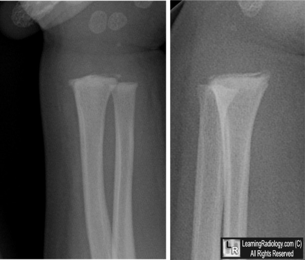
Child abuse. Frontal and oblique views of the radius demonstrate multiple fractures present at the metaphysis of the distal radius (white arrows). A history of child abuse was elicited.
For more information, click on the link if you see this icon 
For this same photo without the annotations, click here
Dahnert 5th edition
Requisites-Pediatric
Radiology
Requisites-Musculoskeletal Radiology
|
|
|

