|
Chance Fracture
Originally most often caused by seat belts as hyperflexion injuries in automobile accidents
o With lap belts, it is now seen more often with falls
· Seat belt injuries usually involve the lower thoracic and upper to mid lumbar spine (L1 and L2 most commonly)
· Chance fractures are hyperflexion injuries in which there is distraction of the posterior elements and impaction of the anterior components of the spine
o Compression component from hyperflexion is usually minor compared to distraction component
· Clinical findings
o Back pain is the most common symptom
o Ecchymosis of anterior abdominal wall should raise suspicion for the presence of this kind of fracture
o Up to 50% of individuals with Chance fractures may also have serious blunt injury to internal organs
§ Injuries involve primarily the pancreas, duodenum and mesentery
§ The same mechanism of injury may not produce a fracture in children but may still be associated with intestinal and urinary bladder injuries
o Since the spinal cord ends at T12-L1, injuries to cord are infrequent but the spinal nerves may be injured resulting in bowel and bladder signs
§ Those with a kyphosis of less than 15° have a better neurologic prognosis
o Mortality is the result of associated internal injuries
· Imaging findings
o CT is the most sensitive study although conventional radiographs are usually obtained first
o Horizontal fracture through the spinous process, laminae, pedicles and vertebral body
o Vertebral body component may be less prominent than the distraction fractures of the posterior elements which are always present
§ Vertebral body component may not be visible

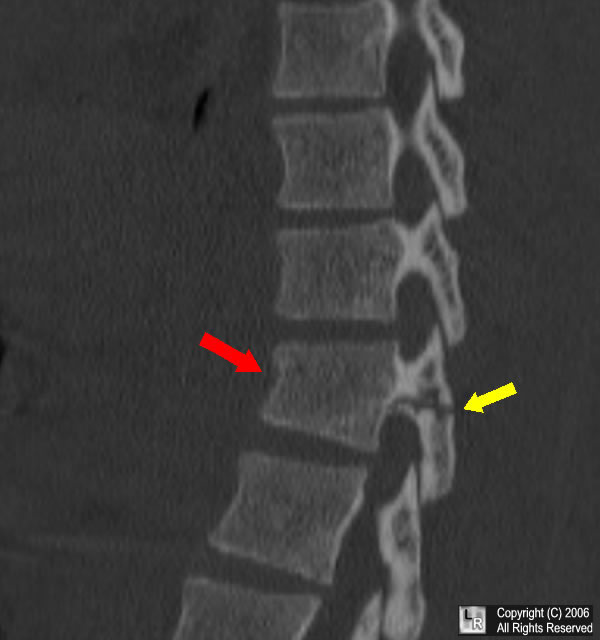
Chance Fracture. Reformatted sagittal CT of the lower thoracic spine demonstrates
a horizontal fracture through the spinous process and pedicles (yellow arrow) and a compression
fracture of the vertebral body (red arrow) characteristic of the hyperflexion distraction-impaction
injury associated with lap seat belt injuries and falls. |
For a photo of the same image without the arrows, click here
o There may be associated soft tissue swelling
o Open pedicle sign – lucency on the medial aspects of the pedicles
o Posterior ligamentous complex tears
§ Up to 50% have injuries to
· Interspinous ligament
· Ligamentum flavum
· Facet capsule
· Posterior annulus
· Thoracodorsal fascia
· Treatment
o Most Chance fractures are managed with immobilization
o Instability is frequently associated with a kyphosis of 20° or more and a kyphosis of 30° or more usually requires internal stabilization
o Main treatment for unstable fractures is surgical fixation with spinal canal decompression

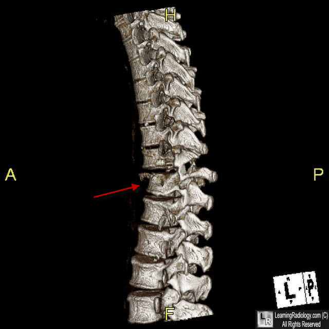
Chance fracture of T10. 3D Reformatted image shows horizontal fracture through T10.
|