RecognizingFractures(and describing them)
© William Herring, MD, FACR
Many fracture examples are part of Radiology Resource and Review (R3) ©2001 Medical College of GeorgiaAll copyrighted images retain the rights of the original copyright holders
Definitions
Fracture
Dislocation
Subluxation
Fracture
A disruption in allor part of thecortex of a bone
All = complete
Part = incomplete =children
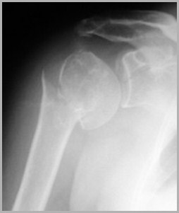
Complete fracture of the surgicalneck of the humerus
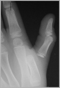
Dislocation
The bonycomponents of a jointno longer are incontact with eachother
There is completedisruption of the joint
R3
Dislocation of the metacarpal-phalyngeal joint of the thumb
Subluxation
The bonycomponents of ajoint are partially incontact with eachother
There is partialdisruption of thejoint
R3
Incomplete Fractures
Greenstick –fracturethrough onecortex
Torus –buckling ofthe cortex
Also called “Buckle fx”
Greenstick Fractures
Buckle Fractures
By the direction of the fracture line
By the relationship of the fragments
By the number of fragments
By communication with the atmosphere
How Fractures Are Described
How Fractures Are Described
By the direction of the fracture line
Transverse
Diagonal or oblique
Longitudinal
Spiral
How Fractures Are Described
By the direction ofthe fracture line
Transverse –perpendicular to thelong axis of the bone
Caused by a forceperpendicular to shaft
How Fractures Are Described
By the direction of thefracture line
Diagonal or oblique –caused by a forceusually applied in thesame direction as thelong axis of the bone
How Fractures Are Described
By the direction of thefracture line
Longitudinal – along thelong axis of the bone

R3
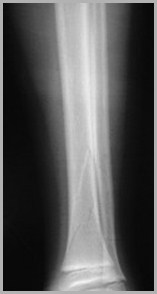
How Fractures Are Described
By the direction of thefracture line
Spiral – a twistingfracture caused by atorque injury such asplanting the foot in ahole while running
How Fractures Are Described
By the relationship of one fracturefragment to another
Displacement
Angulation
Shortening
Rotation
Most fractures display more than one ofthese abnormalities of position
How Fractures Are Described
By convention, abnormalities of positiondescribe the relationship of the distalfracture fragment relative to the proximalfragment
Displacement
The amount of off-set ofthe distal fracturefragment relative to theproximal
There is lateraldisplacement of thedistal femoral fracturefragment in this case
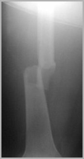
Lateral
Medial
Angulation
The angle away from thenormal that the distalfragment makes with theproximal
In this case the distalfragment is angulatedmedially from theposition it would havewere it not fractured
Lateral
Medial
Shortening
Overlapping of theends of the fracturefragments
Shortening is usuallydescribed by thenumber of centimetersof overlap
There is also medial displacement
Lateral
Medial
Rotation
Almost always involveslong bones (humerusand femur)
Knee joint is in APposition (pointsforward) but anklepoints lateral, in thiscase
Lateral atankle
Anteroposteriorat knee
How Fractures Are Described
By the number offracturefragments
Two fragments -Simple
More than twofragments -Comminuted
Simple
Comminuted
1
2
1
2
3
4
How Fractures Are Described
By the relationshipof the fracture to theatmosphere
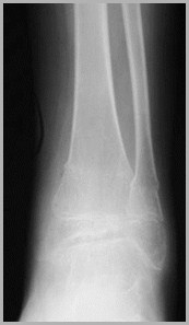
Closed
Open or compound
Best evaluatedclinically
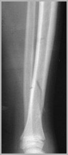
Open or compound fracture of the tibiacommunicates with outside air
Salter-Harris Fractures
Fractures that involve the epiphysealplate alone or in combination with anadjacent part of the bone
Why is the classification important?
Prognostic value
Type I’s and II’s do well
Type IV’s and V’s can develop early fusion ofepiphysis and shortening of that bone
Salter-Harris Classification
Type I – Epiphyseal plate alone
Type II – Epiphyseal plate and metaphysis
Type III – Epiphyseal plate and epiphysis
Type IV – Epiphyseal plate, metaphysisand epiphysis
Type V – crush fracture of epiphyseal plate
Salter-Harris Classification
Type I – Fracturethrough theepiphyseal platealone
Often difficult todetect withoutother side forcomparison
R3
Salter-Harris Classification
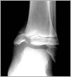
Type II – fracture of themetaphysis and theepiphyseal plate
Most common type ofSalter fracture
“Corner-sign” – smallmetaphyseal fragment
R3
Salter-Harris Classification
Type III – Fractureof the epiphysealplate and theepiphysis
R3
Salter-Harris Classification
Type IV – Fracturethrough themetaphysis,epiphyseal plate andthe epiphysis
Poorer prognosis –i.e. premature closureof epiphysis
R3
Common Fracture Eponyms
Colle’s fracture
Smith’s fracture
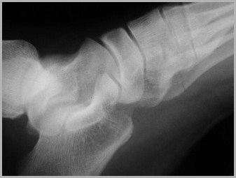
Jones’ fracture
Boxer’s fracture
Common Fracture Eponyms
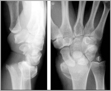
Colle’sfracture –fracture ofthe distalradius withdorsalangulation
R3
Common Fracture Eponyms
Smith’s fracture –fracture of thedistal radius withpalmarangulation
Fall on a flexedhand
R3
Common Fracture Eponyms
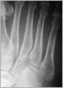
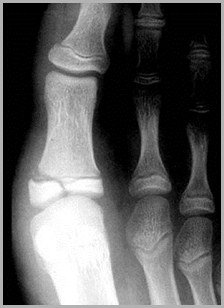
Jones’ fracture – fx base 5th metatarsal
Common Fracture Eponyms
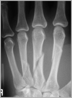
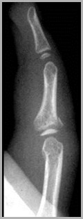
Boxer’s fracture – fxhead 5th metacarpalwith palmarangulation
Most often the resultof punching aperson or wall
R3
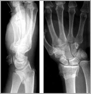
Easily Missed Fractures
Scaphoid fractures
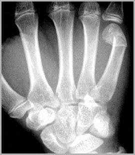
Buckle fractures of radius/ulna
Radial head fracture
Supracondylar fractures in children
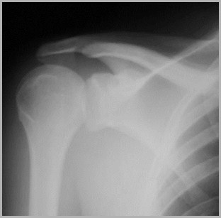
Posterior dislocation of the shoulder
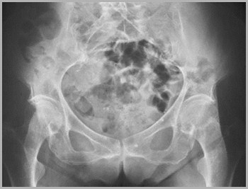
Hip fractures
Easily Missed Fractures
Scaphoid fractures– common
Pain in anatomicalsnuff box
Fall on outstretchedhand
Can lead toavascular necrosis
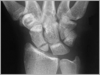
R3
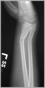
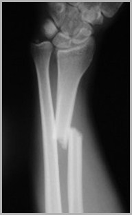
Buckle fractures ofradius/ulna
Children
Look for angulationof cortex
Heal quickly
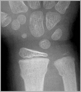
R3
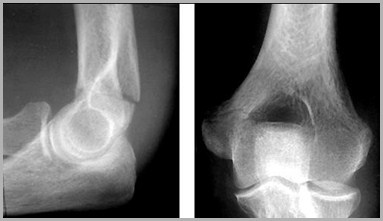
Easily Missed Fractures
Radial headfracture
Common
May requiremultiple viewsto see it
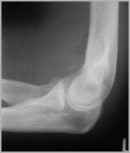
Easily Missed Fractures
Supracondylarfractures inchildren
Easily Missed Fractures
R3
Supracondylar fractures can be difficult– especially in children (this is an adult)
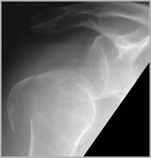
Posteriordislocation of theshoulder
Humeral head lookslike “lightbulb”
Usually needanother view likeaxillary or Y view
Easily Missed Fractures
R3
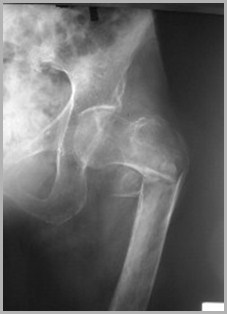
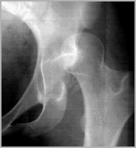
Hip fractures
May be very subtleand require bonescan or MRI fordiagnosis
In this case, whitezone of sclerosis isan impactedsubcapital fracture
Easily Missed Fractures
R3
Jones’ fracture –fracture of the baseof the 5th metatarsal
Avulsion typefracture frequentlycaused by pull ofperoneus brevistendon
Easily Missed Fractures
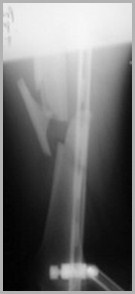
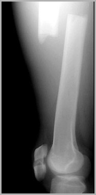
Fracture Healing
Indistinctness of fracture line
Bony callous production
Bridging of fracture
Obliteration of fracture line
Remodeling of bone
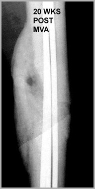
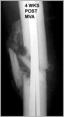
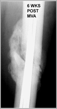
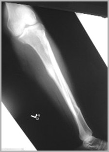
Series of images shows indistinctness of fracture line with bony callousformation and, finally, obliteration of the fracture line. There are metallic rodstransfixing the fracture.
R3
Quiz
Correct answers in red on slide after question
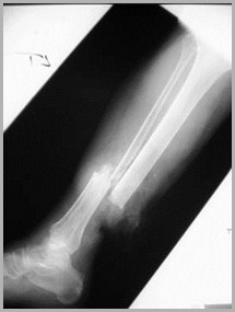
The two terms below thatbest describe theposition of these femoralfracture fragments are:
1.Angulated
2.Comminuted
3.Displaced
4.Shortened
5.Rotated
There is also a fracture of the patella
The terms below that bestdescribe this fractureare:
1.Angulated
2.Comminuted
3.Rotated
4.Greenstick
5.Spiral
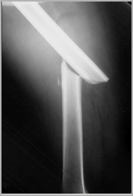
This is an example of a:
1.Comminuted fracture
2.Dislocation
3.Displaced fracture
4.Subluxation
Posterior dislocation of the hip
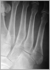
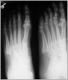
This fracture most likelyoccurred:
1.An hour ago
2.A day ago
3.A week ago
4.A decade ago
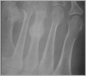
Healing “stress-type” fracture of the 3rdmetatarsal already has significantcallous formation indicating it is atleast several days old
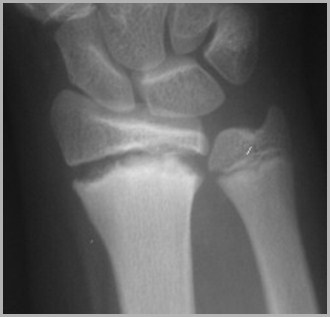
This is an example of aSalter-Harris:
1.Type I fracture
2.Type II fracture
3.Type III fracture
4.Type IV fracture
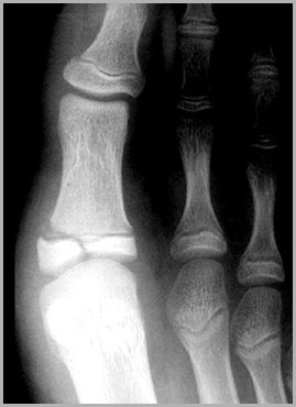
There is a fracture that extends throughthe epiphysis but not the metaphysis.There is also a fracture of theepiphyseal plate.