


RadiographicInterpretation in theMediastinumRadiographicInterpretation in theMediastinum
Adam Guttentag M.D.Adam Guttentag M.D.
MediastinumMediastinum
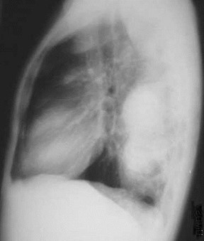
Radiologic anatomyRadiologic anatomy
CXRCXR
CTCT
Normal or not?Normal or not?
Compartments and their pathologies:Compartments and their pathologies:
AnteriorAnterior
MiddleMiddle
PosteriorPosterior
Mediastinal AnatomyMediastinal Anatomy
Bounded laterally by parietal pleura ofeach lungBounded laterally by parietal pleura ofeach lung
Superior margin is thoracic inletSuperior margin is thoracic inlet
Defined by plane of 1st ribsDefined by plane of 1st ribs
Inferior margin is diaphragmInferior margin is diaphragm
Tissue planes extend superiorly intoneck around great vessels, trachea andesophagusTissue planes extend superiorly intoneck around great vessels, trachea andesophagus
Connects to abdominal cavity viaesophageal, caval and aortic hiatusesConnects to abdominal cavity viaesophageal, caval and aortic hiatuses
Connects to lungs along hilar vesselsand bronchiConnects to lungs along hilar vesselsand bronchi
Radiologic AnatomyRadiologic Anatomy
Landmarks to look for onthe chest radiograph:Landmarks to look for onthe chest radiograph:
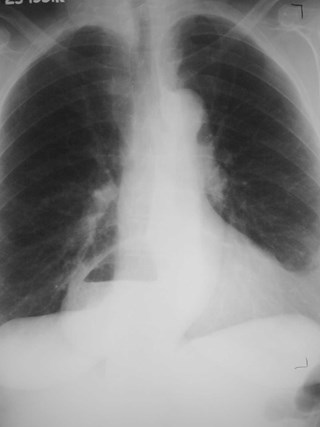
TracheaTrachea
Paratracheal stripeParatracheal stripe
Junction linesJunction lines
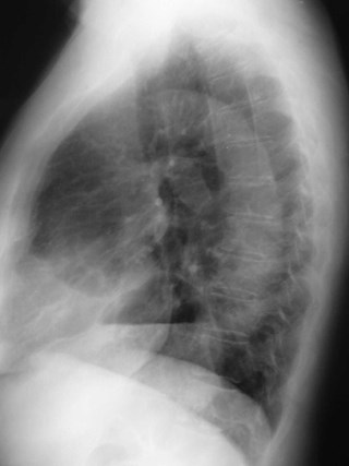
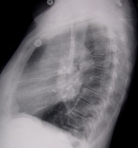
Descending aortic contourDescending aortic contour
Azygo-esophageal contourAzygo-esophageal contour

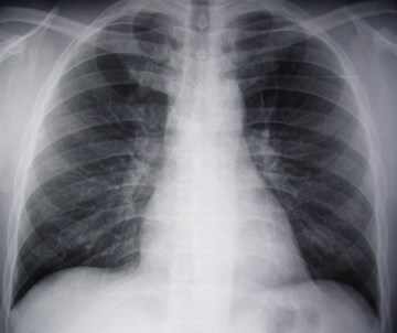
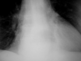
Tracheal air column
Should be straightShould be straight
Slight bend toright due to aorticarch on its left.Slight bend toright due to aorticarch on its left.
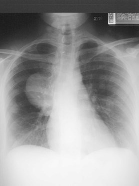
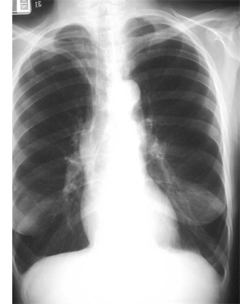
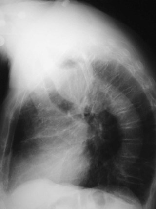
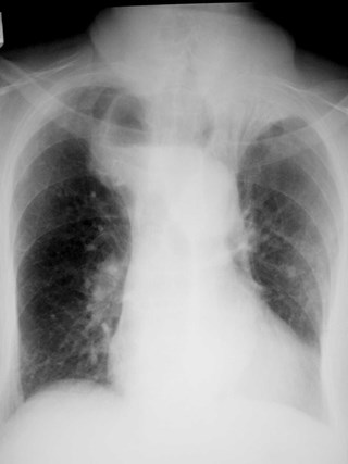
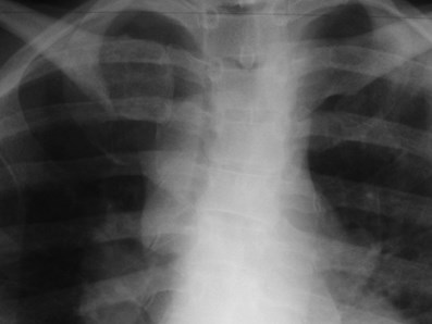
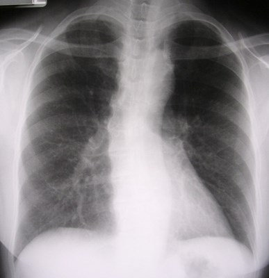
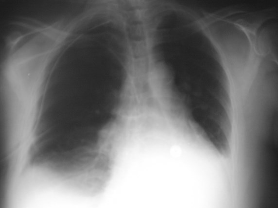
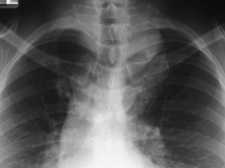
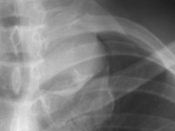
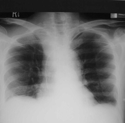
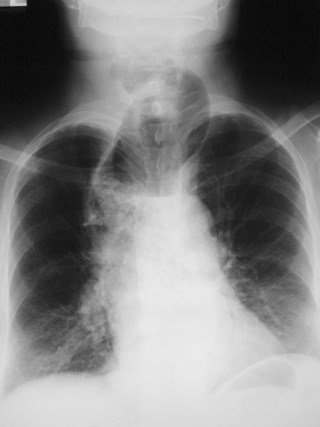
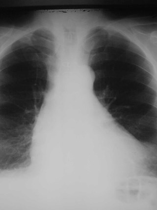
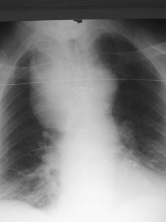
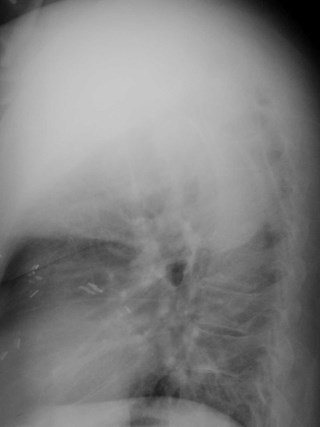
Right Aortic ArchRight Aortic Arch
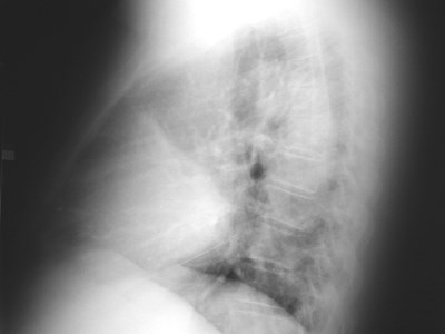
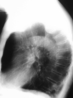
70 yo with dysphagia70 yo with dysphagia
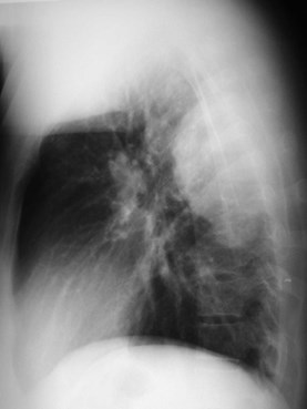
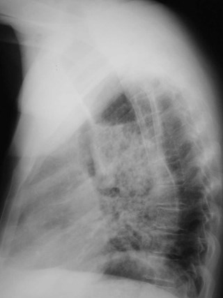
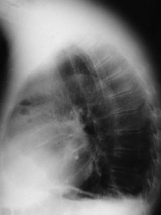
Esophageal CarcinomaEsophageal Carcinoma
Zenker DiverticulumZenker Diverticulum
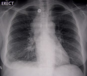
Erect CXR

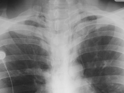
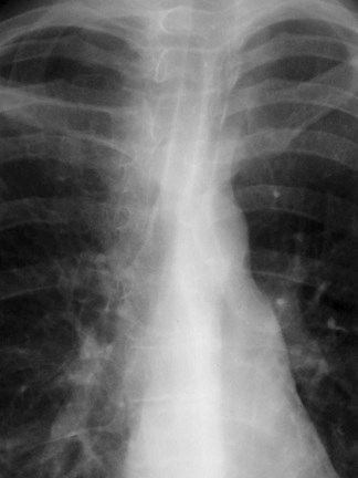
Right paratracheal stripe
Extends fromthoracic inlet toazygos vein
Widens out atazygos vein
No more than 3-4mm thick
SarcoidSarcoid

Old film
Hodgkins diseaseHodgkins disease

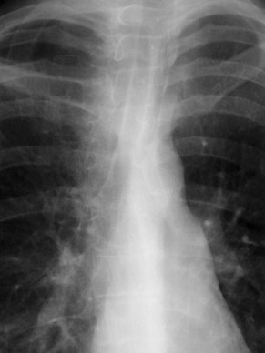
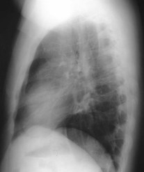
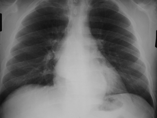
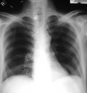
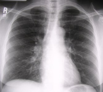
Aortic ContourAortic Contour
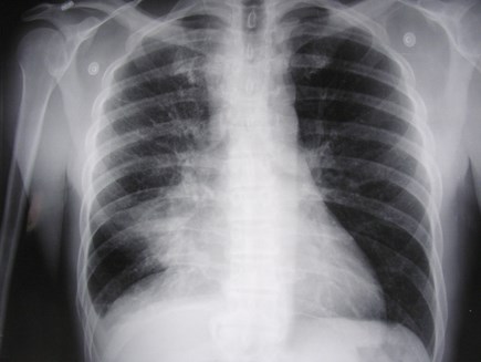
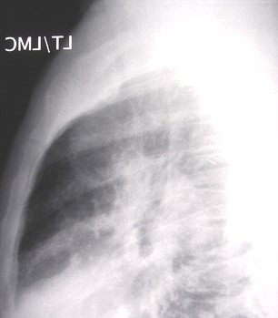
HoarseHoarse
One year ago
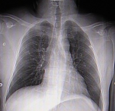
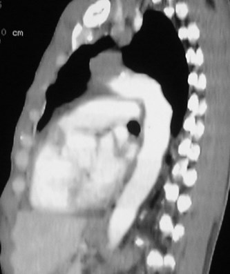
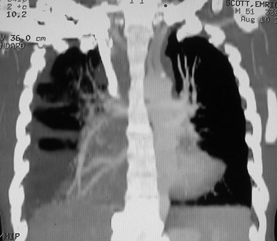
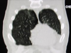
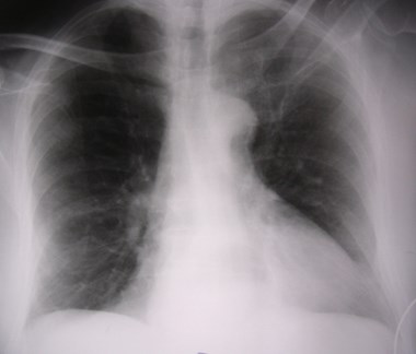
Lung CancerLung Cancer
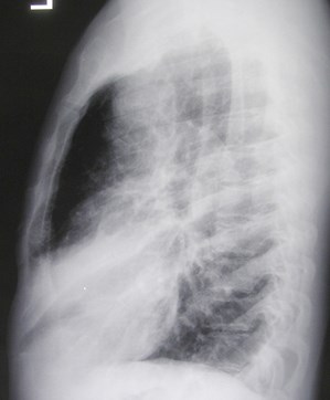
Chronic CoughChronic Cough

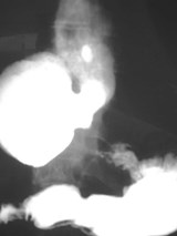
Azygo-esophageal recessAzygo-esophageal recess
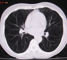
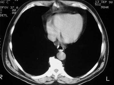
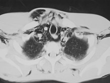
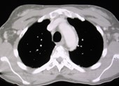
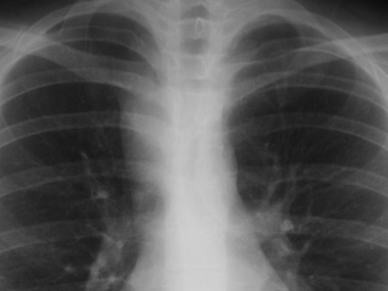
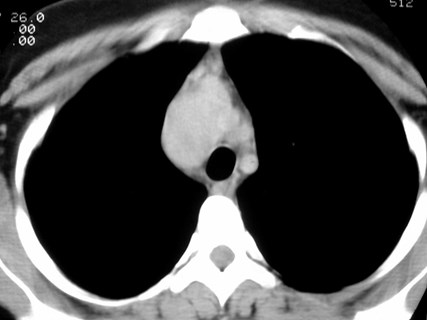
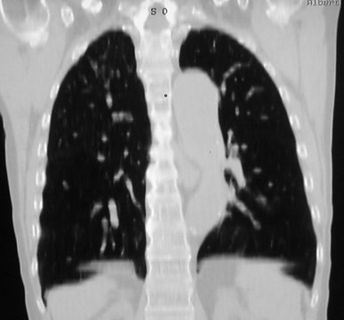
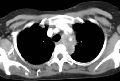
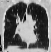
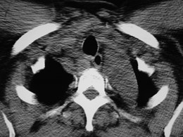
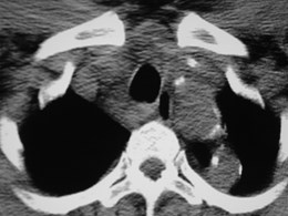
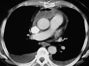
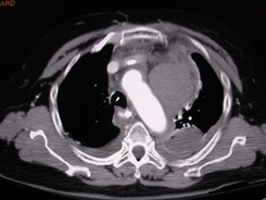
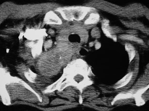
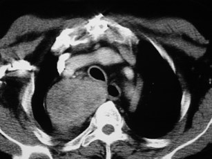
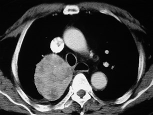
Bronchogenic CystsBronchogenic Cysts
Anterior junction lineAnterior junction line
Extends from thoracic inletinferiorly and to the leftExtends from thoracic inletinferiorly and to the left
Widens superiorlyWidens superiorly

Anterior PneumothoraxAnterior Pneumothorax
Posterior Junction LinePosterior Junction Line
Not always seenNot always seen
Usually in emphysemaUsually in emphysema
Extends inferiorly to thelevel of the aortaExtends inferiorly to thelevel of the aorta
Widens superiorly tooutline apices of lungsWidens superiorly tooutline apices of lungs
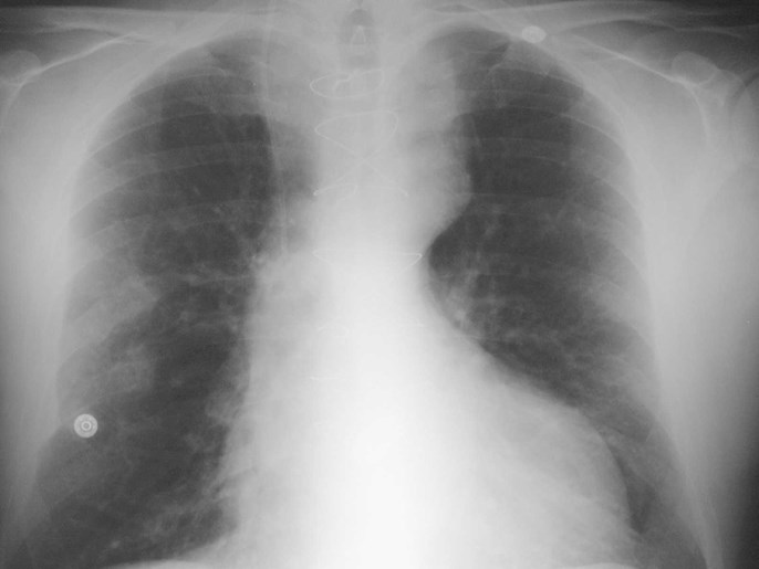
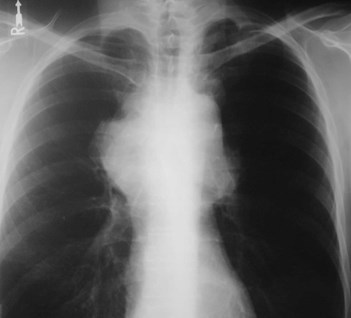
Wide Mediastinum???Wide Mediastinum???
First ask…First ask…
Is it technical?Is it technical?
PositioningPositioning
rotated:?rotated:?
AP?AP?
Lordotic?Lordotic?
Supine?Supine?
InspirationInspiration
If so, repeat the film before proceeding to CT.If so, repeat the film before proceeding to CT.
Then ask…Then ask…
Is it new?Is it new?
GET OLD FILMS before you get a CT scan unless timeis critical..GET OLD FILMS before you get a CT scan unless timeis critical..
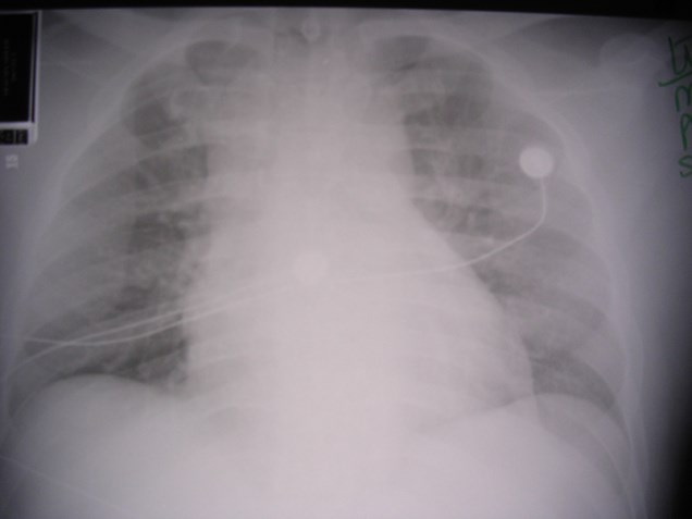
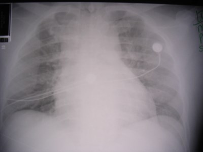
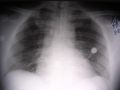
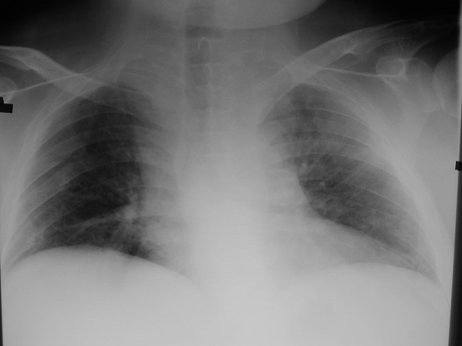
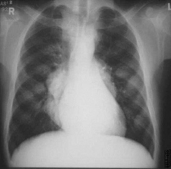
30 year old man in motor vehicle accident
AP supine
AP upright
PA upright
Think about normal variants!Think about normal variants!
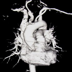
Vascular structuresVascular structures
Aortic anomaliesAortic anomalies
Tortuous vesselsTortuous vessels
Azygos veinAzygos vein
LipomatosisLipomatosis
Fat padsFat pads
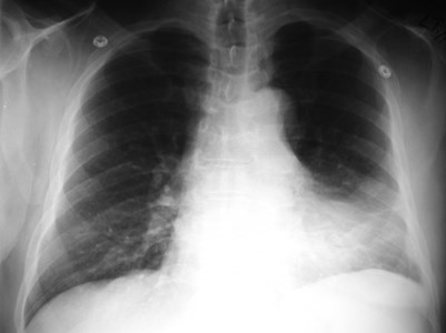
Severe asthma on longterm steroids
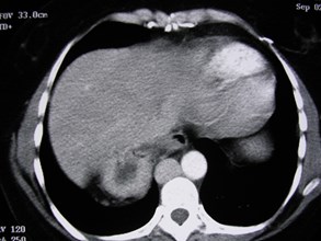
Mediastinal Lipomatosis
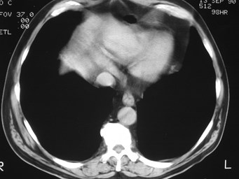
Pericardial Fat Pad
Tortuous brachiocephalic vesselsTortuous brachiocephalic vessels
Azygos continuation of the IVCAzygos continuation of the IVC
Apical Aortic ArchApical Aortic Arch
Check the old film!Check the old film!
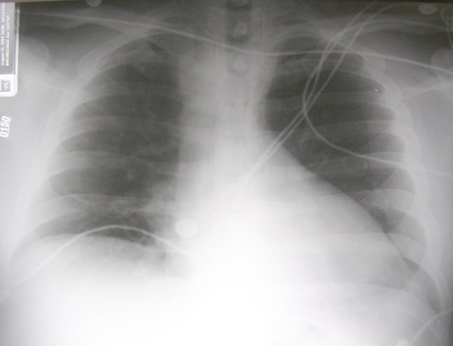
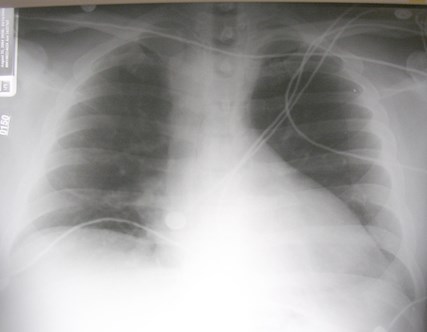
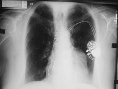
Recent CVC stickRecent CVC stick

2 days earlier
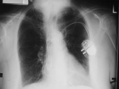
One year earlier
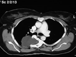
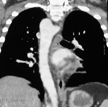
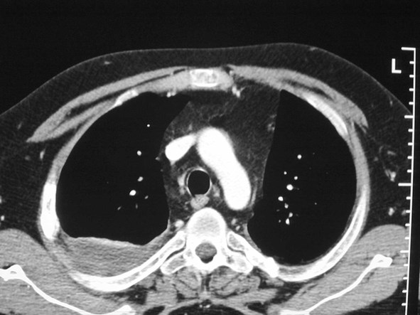
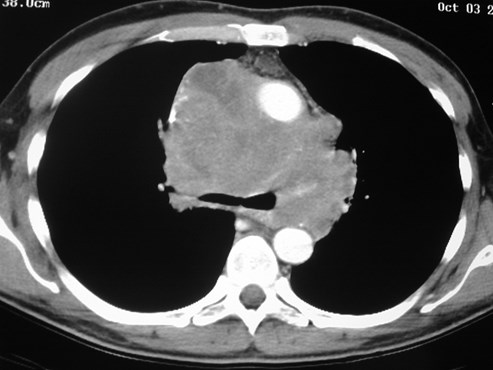
Pericardial LymphomaPericardial Lymphoma
Special case:Air fluid levelsSpecial case:Air fluid levels
Think bowel or abscess:Think bowel or abscess:
Hiatal herniaHiatal hernia
Zenker diverticulumZenker diverticulum
Epiphrenic diverticulumEpiphrenic diverticulum
AchalasiaAchalasia
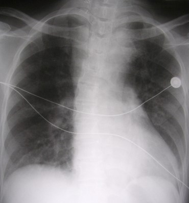
Mediastinal abscessMediastinal abscess
AchalasiaAchalasia
Hiatal herniaHiatal hernia

Epiphrenic diverticulumEpiphrenic diverticulum
Mediastinal abscess after CABGMediastinal abscess after CABG
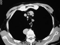
Mediastinal CompartmentsMediastinal Compartments
Arbitrary division of contiguous spaces,especially anterior and middle compartmentsArbitrary division of contiguous spaces,especially anterior and middle compartments
Useful in grossly narrowing differential diagnosisUseful in grossly narrowing differential diagnosis
Often significant crossing of compartmental“boundaries”, making classification difficult.Often significant crossing of compartmental“boundaries”, making classification difficult.
Anterior mediastinumAnterior mediastinum
Space anterior to greatvessels and heart,behind the sternumSpace anterior to greatvessels and heart,behind the sternum

Anterior mediastinumAnterior mediastinum
Most common:Most common:
Thymic lesionsThymic lesions
Germ cell origin tumorsGerm cell origin tumors
+/- Thyroid masses+/- Thyroid masses
LymphomaLymphoma
MetastasesMetastases
Also:Also:
Foregut cysts, nerve tumors, aortic aneurysm, etc.Foregut cysts, nerve tumors, aortic aneurysm, etc.
Anterior mediastinal massesAnterior mediastinal masses
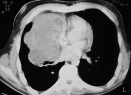
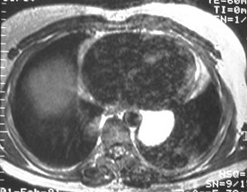
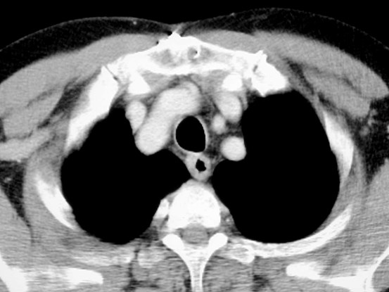
Thymic hyperplasia
teratocarcinoma
teratoma
lymphoma
thymoma
hemorrhage
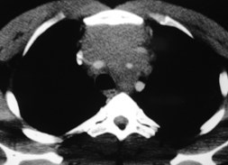
Middle MediastinumMiddle Mediastinum
Contains heart, greatvessels, esophagus,trachea, lymph nodes,nerves.Contains heart, greatvessels, esophagus,trachea, lymph nodes,nerves.
CXR abnormality inparatracheal area,azygo-esophagealrecess, retrocardiac areaCXR abnormality inparatracheal area,azygo-esophagealrecess, retrocardiac area
May be difficult to seeon lateral viewMay be difficult to seeon lateral view

Middle MediastinumMiddle Mediastinum
Lymph nodesLymph nodes
LymphomaLymphoma
MetastasesMetastases
Foregut cystsForegut cysts
Vascular lesions e.g. aneurysmVascular lesions e.g. aneurysm
Cardiac abnormalitiesCardiac abnormalities
Bronchogenic carcinomaBronchogenic carcinoma
Hiatal hernia, other esophageal or GI lesionsHiatal hernia, other esophageal or GI lesions
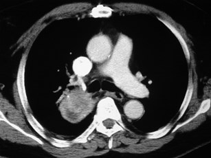
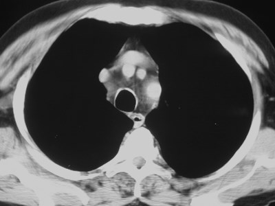
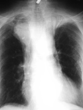
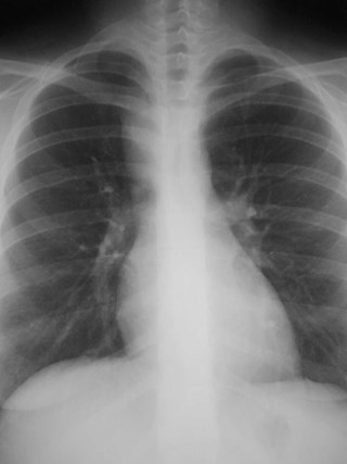
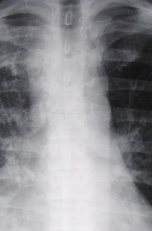
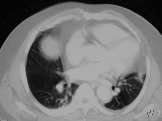
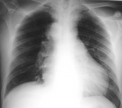
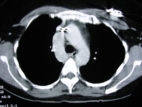
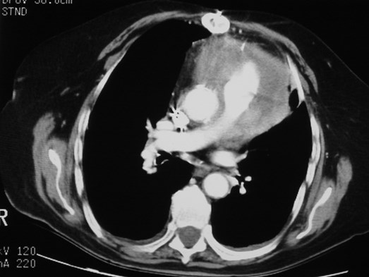
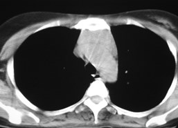
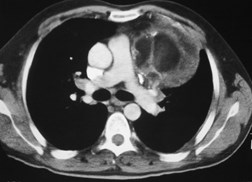
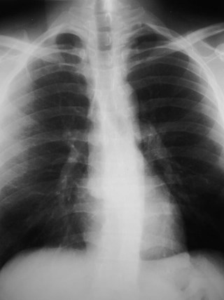
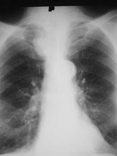
Mediastinal GoiterMediastinal Goiter
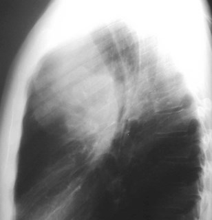
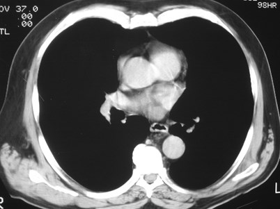
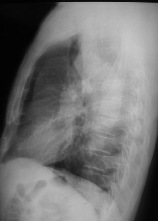
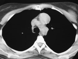
Small Cell Lung CaSmall Cell Lung Ca
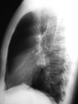
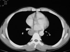
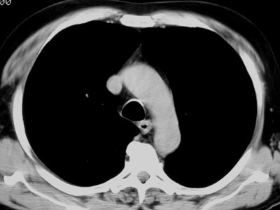
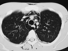
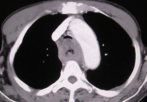
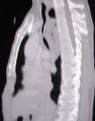
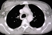
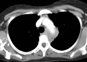
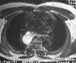
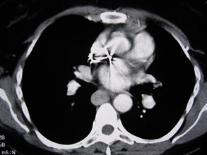
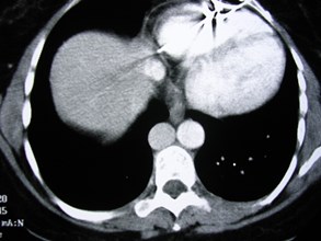
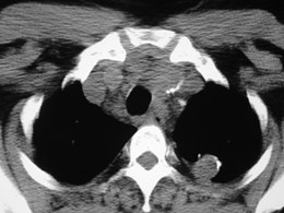
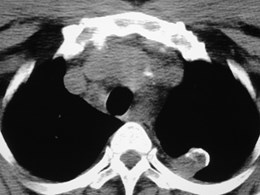
Bronchogenic CystBronchogenic Cyst
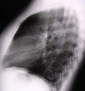
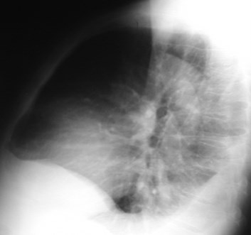
Posterior mediastinumPosterior mediastinum
Paraspinal areaParaspinal area
Masses usually visible onlateral film overlying spineMasses usually visible onlateral film overlying spine
May obscure descendingaorta contourMay obscure descendingaorta contour
Posterior MediastinumPosterior Mediastinum
Neurogenic tumorsNeurogenic tumors
SchwannomaSchwannoma
Sympathetic nerve tumors e.g.ganglioneuromaSympathetic nerve tumors e.g.ganglioneuroma
Neurenteric cyst, lateralmeningoceleNeurenteric cyst, lateralmeningocele
Lymph nodes (unusual asonly area involved)Lymph nodes (unusual asonly area involved)
ExtramedullaryhematopoesisExtramedullaryhematopoesis
Vertebral tumors, spursVertebral tumors, spurs

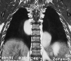
SchwannomaSchwannoma
Extramedullary HematopoesisExtramedullary Hematopoesis
Lateral Meningocele in NF-1Lateral Meningocele in NF-1