
Other First TrimesterIssues
Retained products ofconception
Gestational trophoblasticneoplasia
Puerperal infections
Uterine and adnexal masses
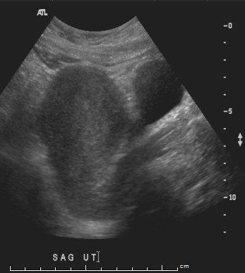
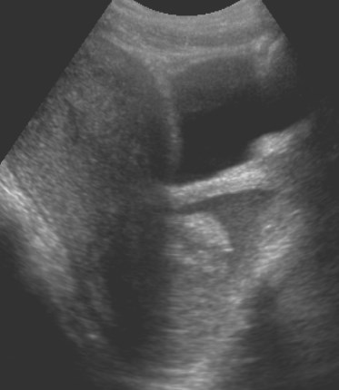
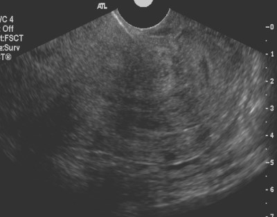
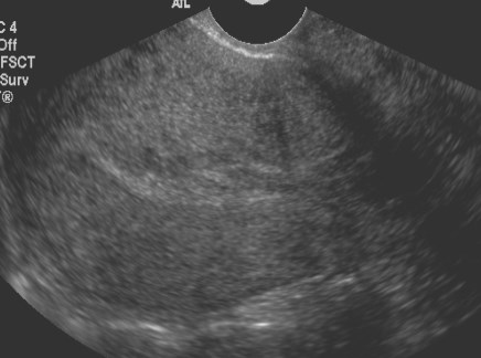
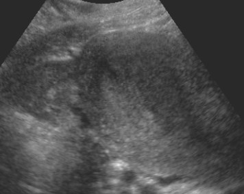
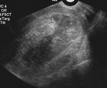
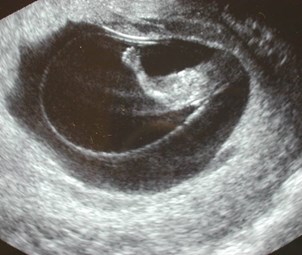
S/P therapeutic abortion one weekearlier with persistent bleeding
Retainedproducts ofconception
Retained Products of Conception
Retained Products ofConception
Very few large studies, small amountsmay be clinically insignificant
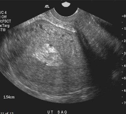
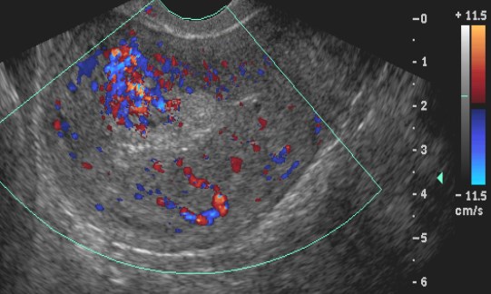
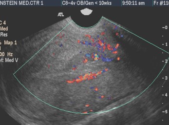
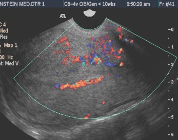
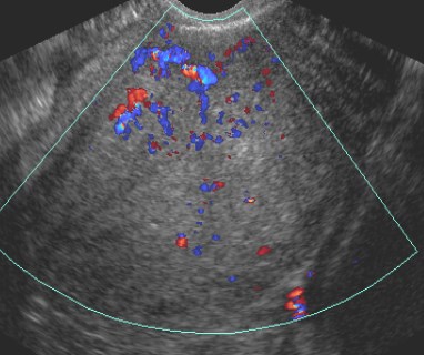
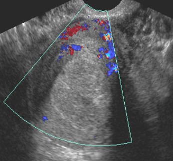
Focal echogenic mass most usefulfinding
Unlikely with thin endometrium (8-10mm)
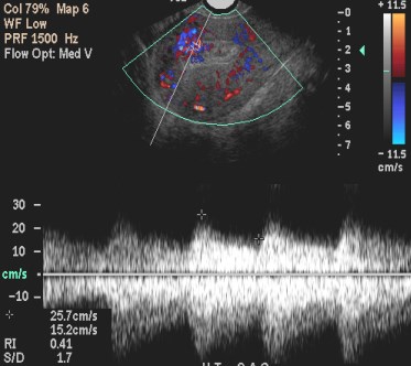
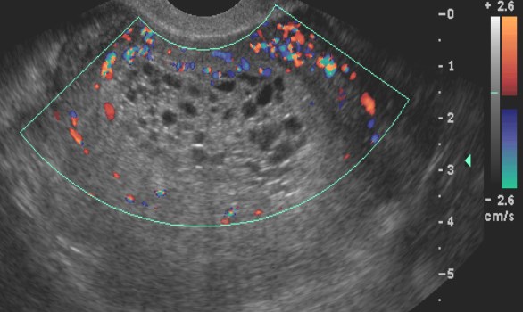
Focal low resistance flow ischaracterisitic, but may be absent withnecrotic placenta
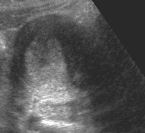
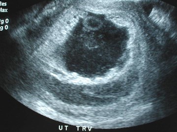
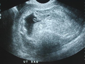
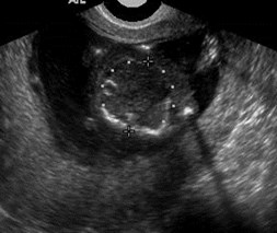
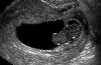
Patient with history of 3 months gestation,now with heavy bleeding, passing tissue
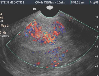
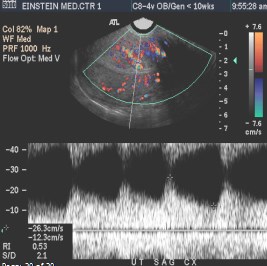
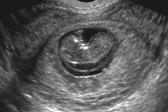
Spontaneous abortionwith clots and blood invagina and small focusretained products ofconception
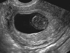
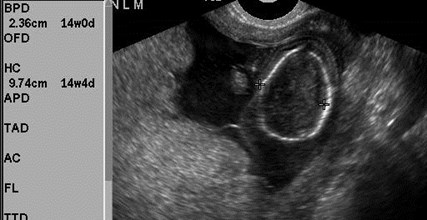
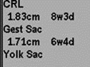
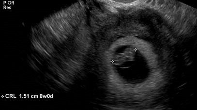
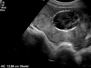
Definitive 7 week IUP, patientwent for therapeutic abortion
Trv
Sag right
Sag left
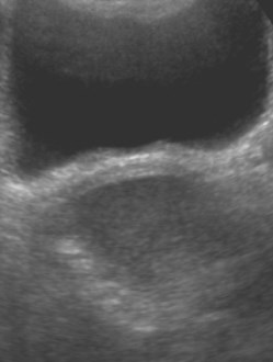
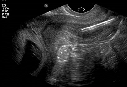
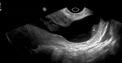
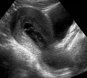
3 days after abortion withnew, heavy bleeding, giveshistory of uterine anomaly
Sag R
SagL
Completed abortion right horn ofbicornuate uterus with bleeding due toshedding of decidua in smaller left horn
Gestational TrophoblasticDisease
Abnormal proliferation of pregnancyassociated trophoblastic disease withmalignant potential
Hydatidiform Mole: benign
Invasive Mole and Choriocarcinoma-malignant
Placental site trophoblastic tumor is rareform of GTD often with normal or lowHCG
Atypical molar pregnancy- appearancesimilar to incomplete abortion

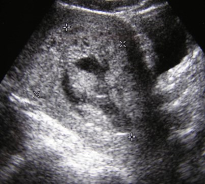
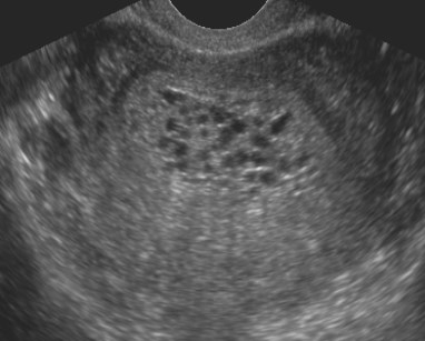
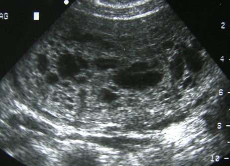
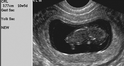
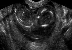
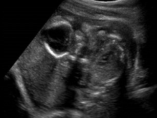
First trimester molar pregnancy
HCG 181,000
Molar Pregnancy
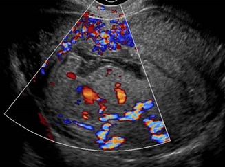
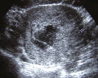
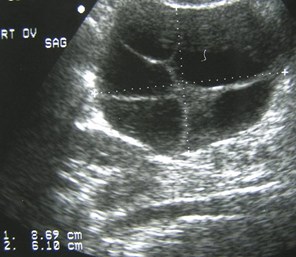
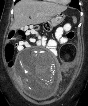
14 weeks by dates, bleeding
Retained placenta, residual sac
Retained sac and hydropic placenta
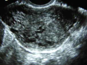
Bleeding HCG 956
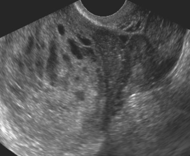
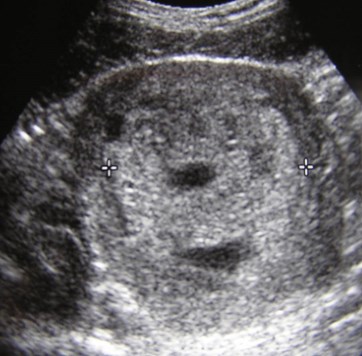
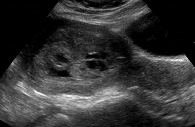
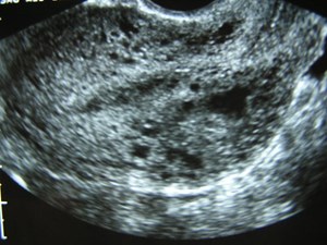
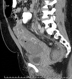
Pregnant with bleeding
Molar Pregnancy
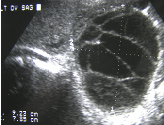
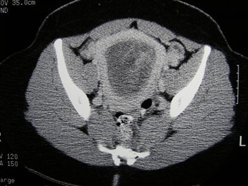
Second trimester molar pregnancywith bilateral theca lutein cysts
Hydatidiform Mole
Most common form of gestationaltrophoblastic disease 1/1000- 1/2000pregnancies, most frequently in teens and 40s
First trimester appearance variable: sac-likecollection, complex echogenic mass, thickendometrium
Only 50% correctly diagnosed prospectivelycompared to 100% of 2nd trimester molarpregnancies
Lazarus, JUM 1997
Hydatidiform Mole
Two forms: complete and partial
Complete Mole
–46XX, all from sperm, fertilization of single egg with lostchromosomes, embryo dies very early
–Hydropic degeneration and absence of vessels in swollenvilli
–Theca lutein cysts 2° overstimulation of lutein elements byelevated HCG
Partial Mole
–Usually 69XXY, due to fertilization of one egg by two sperm
–Embryo lives for several weeks
Lazarus, JUM 1997

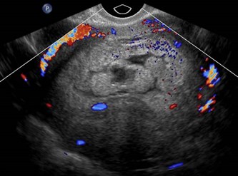
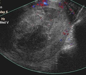
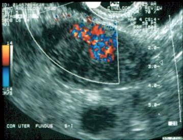
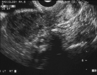
History molar pregnancy with rising HCG
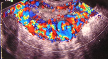
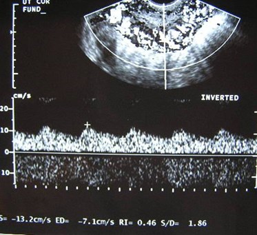
Invasive Mole
Gestational TrophoblasticTumor
Invasive Mole: persistent trophoblasticproliferation with penetration intomyometrium and other local tissues,metastases are rare
50% after hydatidiform mole
25% after abortion
25% after normal or ectopic pregnancy
Choriocarcinoma: absence of villous pattern,early metastases with 75% to lungs, alsovagina, brain, liver, kidneys, ovaries, bowel
Green etal. Radiographics 1996; 16:1371
Diffuse Lymphedema, XO
Diffuse Lymphedema, XO
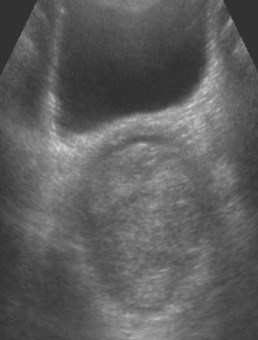
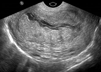
Uterine Synechiae
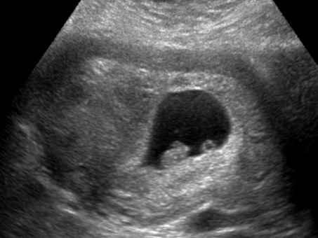
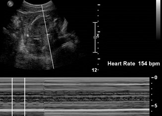
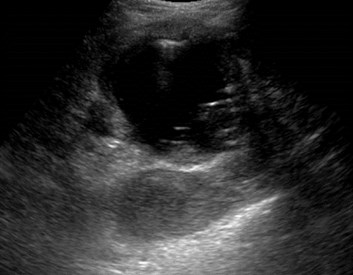
Negative cardiac activity
8 week IUP with IUD in cervix
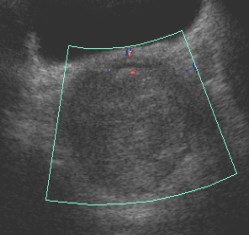
Passing clots
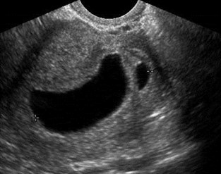
Inevitable AB- dilated cervixcontaining gestational sac
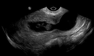
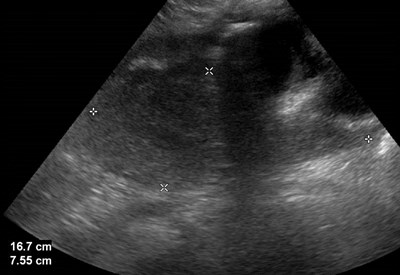
31 week pregnancy, recurrent L sided pain,normal US @ 8 weeks
Normal pregnancy, intermittent torsionleft cystic teratoma
Oligohydramnios
Secondary to ruptured membranes
Postpartum Complications
Infections
–Septic pelvic thrombophlebitis
–Parametrial phlegmon
–Pelvic abscess
–Uterine dehiscence
Retained placenta
Hematoma: bladder flap hematomas inlower anterior uterine segment, region> 2-3 cm
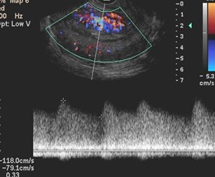
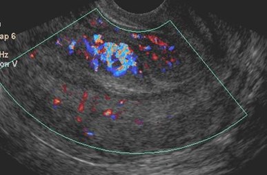
Uterine Arteriovenous Malformation
Brown U Quarterly 2005; 21:27
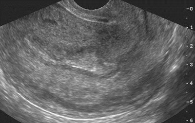
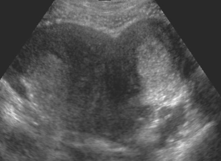
Normal postpartum uterus
Uterus decreases to non-gravidsize by 6-8 weeks postpartum
Transabdominal imaging oftenquite adequate, especially inimmediate post partum period
Endometrium: fluid, clots, smallfoci of air for few weeks, overlapswith appearance of endometritis
Fever and bleeding one week after delivery
Clot in uterus, clinicaldiagnosis of endometritis
Fever after C-section
Infected hematomain C-section scar
Fever after C-section
Infected C-section hematoma
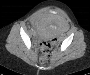
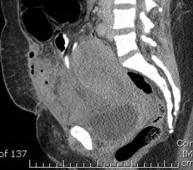
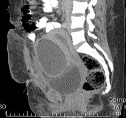
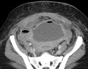
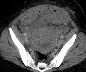
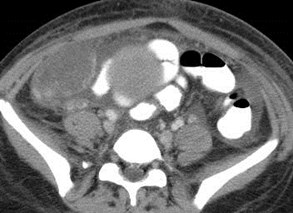
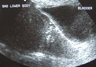
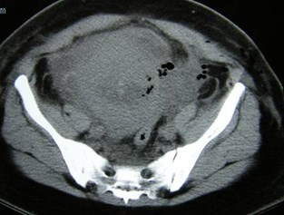
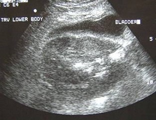
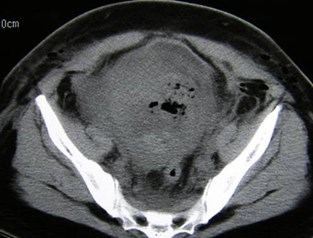
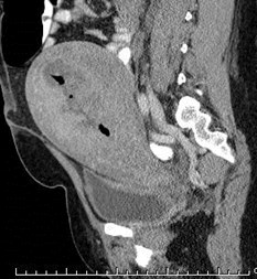
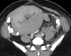
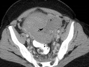
Hx placenta accreta, fever
Abscess and RPOC
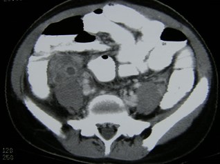
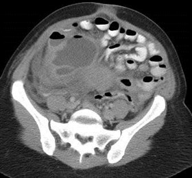
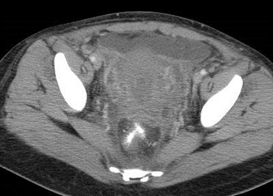
Fever after delivery
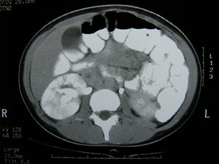
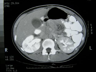
Right ovarian vein thrombosisand right pyelonephritis
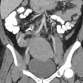
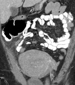
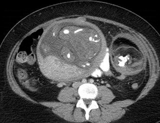
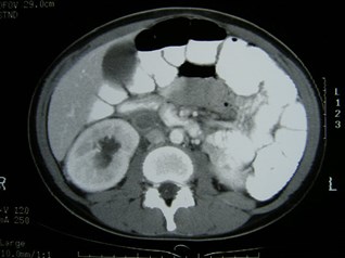
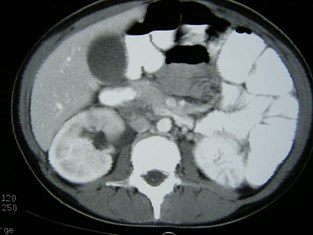
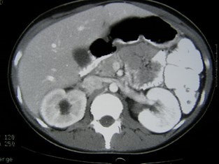
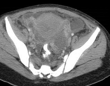
RLQ pain, 9w IUP
R ovarian vein thrombosis, normal appendix
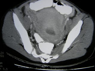
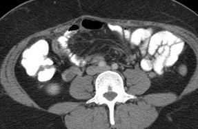
Fever after delivery
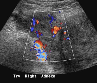
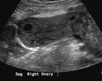
Diffuse, septic pelvicthrombophlebitis
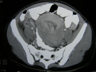
Fever, several days after C-section
Endometritis and Myometritis-clostridium
Fever and pain, vaginal deliverywith manual extraction of placenta
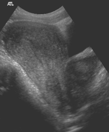
Uterine Dehiscence
Uterine AVM
Case compliments of Dr. Jill Langer, U of P

Bye-bye