
Differential Diagnoses In Heart Disease
© William Herring, MD, FACR
ABCs of Heart Disease
●Pattern recognition approach
●Requires heart to assume adult shape
●Works from 2 years and older
Neonates and Infants
●Heart is globular in shape
●Requires examination of
■Pulmonary vasculature
●Requires knowledge of
■Presence or absence of cyanosis
●Must memorize 4 lists ofDDXs with 5 diagnoses on each
Pattern Recognition in the Newborn
●Even some newborn hearts have acharacteristic shape
●Aunt Minnie, Uncle Murray, CousinAngie and Cardio Cat

Nine Lesions Which Produce 75% of All SevereCongenital Heart Lesions In the Neonate
Decreased flow
1. Tetralogy of Fallot
2. Tricuspid Atresia
3. Severe Pulmonic Stenosis
4. Ebstein’s
Increased Flow
5. Transposition
6. VSD
Nine Lesions Which Produce 75% of All SevereCongenital Heart Lesions In the Neonate
Pulmonary venous hypertension
7. Hypoplastic left heart syndrome
8. Coarctation of the aorta
9. TAPVR with infradiaphragmatic obstruction
What remains
Left-to-right shunts
ASD
PDA
Truncus arteriosus
Cyanotic
Cyanotic
Cyanosis With Decreased Vascularity
Transposition*
Tricuspid atresia*
Truncus-type IV
Tetralogy of Fallot
Ebstein's
* Also appears on DDx of Cyanosis with Vascularity
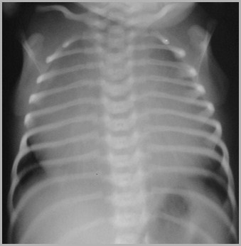
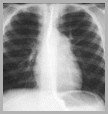
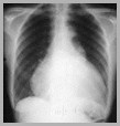
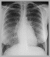
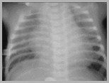
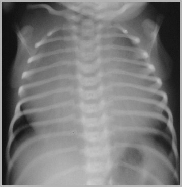
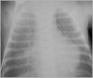
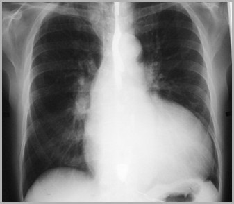
Ebstein’s Anomaly
Ebstein’s Anomaly
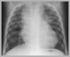
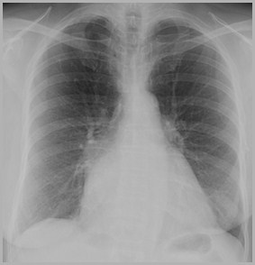
Marked Cardiomegaly at Birth with Cyanosis, Vasculature
Cyanotic
Cyanotic
Cyanosis With Increased Vascularity
Transposition*
Tricuspid atresia*
Truncus types I, II, III
TAPVR
Single ventricle
* Also appears on DDx of Cyanosis with Vascularity
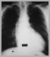
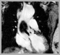
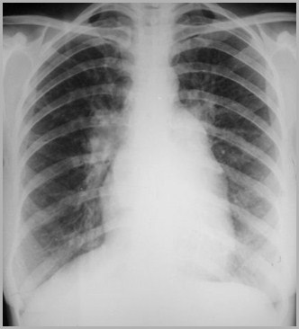
Totalanomalous
venous
return
(TAPVR)
Type 1
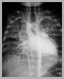
Totalanomalous
venous
return
(TAPVR)
Type 1
Acyanotic

Acyanotic
Cardiomegaly with Normal Vasculature
Viral myocarditis
Endocardial fibroelastosis
Aberrant left coronary artery
Cystic medial necrosis
Diabetic mother
Acyanotic
Acyanotic
Infant of diabetic mother
Infant of diabetic mother
CHF In NewbornImpede Return of Flow to Left Heart
Infantile coarctation
Congenital aortic stenosis
Hypoplastic left heart syndrome
Congenital mitral stenosis
Cor triatriatum
Obstruction to venous return fromlungs
TAPVR from below diaphragm
Hypoplastic left heart syndrome
Hypoplastic left heart syndrome
CHF InChronologic Sequence
CHF InChronologic Sequence
Commonest Cause of CHF In Chronologic Sequence

Time
Cause
< 24 hrs
Commonest Cause of CHF In Chronologic Sequence
Time
Cause
< 24 hrs
Intrauterine Arrhythmia
Commonest Cause of CHF In Chronologic Sequence
Time
Cause
< 24 hrs
Intrauterine Arrhythmia
First week
Commonest Cause of CHF In Chronologic Sequence
Time
Cause
< 24 hrs
Intrauterine Arrhythmia
First week
Hypoplastic LeftHeart Syndrome
Commonest Cause of CHF In Chronologic Sequence
Time
Cause
< 24 hrs
Intrauterine Arrhythmia
First week
Hypoplastic LeftHeart Syndrome
2-6 weeks
Commonest Cause of CHF In Chronologic Sequence
Time
Cause
< 24 hrs
Intrauterine Arrhythmia
First week
Hypoplastic LeftHeart Syndrome
2-6 weeks
Infantile Coarctation
Commonest Cause of CHF In Chronologic Sequence
Time
Cause
< 24 hrs
Intrauterine Arrhythmia
First week
Hypoplastic LeftHeart Syndrome
2-6 weeks
Infantile Coarctation
1-4 months
Commonest Causes of CHF In Chronologic Sequence
Time
Cause
< 24 hrs
Intrauterine Arrhythmia
First week
Hypoplastic LeftHeart Syndrome
2-6 weeks
Infantile Coarctation
1-4 months
Large L R shunts
Common Principles
Common Principles
Principles
Left-to-right shunts → increased flow tolungs
Increased flow to the lungs is 2° left-to-right shunts
Increased flow ≠ pulmonary venoushypertension

ASD
MS
Principles
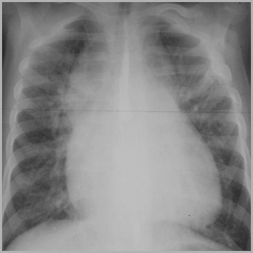
Pulmonary venous hypertension iscaused by CHF or mitral stenosis
Mitral regurgitation does not producepulmonary venous hypertension
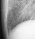
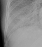
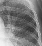
Laminar effusions are 2° pulmonaryvenous hypertension
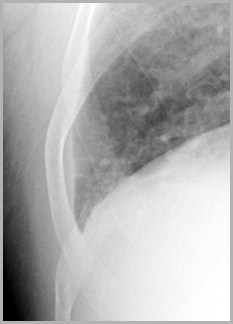
Laminar Effusion
Principles
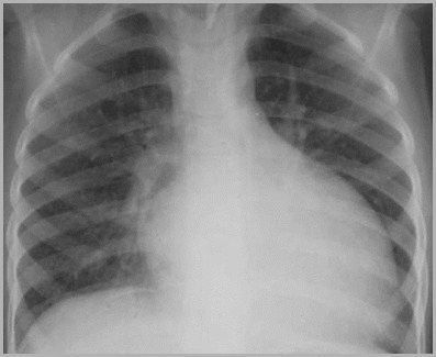
Obstruction to outflow → hypertrophyin ventricles
Obstruction to outflow → enlargementof atria
Regurgitation → larger chambers thanstenosis
Aortic Stenosis
Aortic Regurgitation
Principles
Cardiomegaly is caused by ventricularenlargement
Cardiomegaly is not caused by atrialenlargement
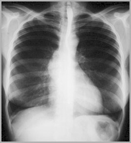
Mitral Stenosis
Principles
Right-to-left shunts produce cyanosis
Rib notching only occurs in the highpressure circuit
Calcification of the PA or left atrium is2° PAH
PrinciplesCHF
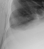
Kerley B lines
Pleural effusions
Fluid in the fissures
Peribronchial cuffing
A
B
C
D
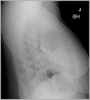
PrinciplesValves
The aortic valve lies over spine andabove the diagonal line on the lateral
The mitral valve lies to left of spine andbelow diagonal line on the lateral
The pulmonic valve is never replaced
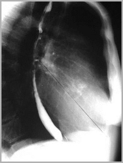
Mitral and Aortic Valves
Patient with prosthetic mitral and aortic valves
A
M
Most Commons
Most common congenital cardiac lesion
1.Bicuspid aortic valve
2.VSD
3.Hypoplastic left heart syndrome
Most common congenital cardiac lesion
1.Bicuspid aortic valve
2.VSD
3.Hypoplastic left heart syndrome
Most common congenital heart lesion(excluding bicuspid aortic valve)
1.Truncus arteriosus
2.Prolapsing mitral valve
3.VSD
Most common congenital heart lesion(excluding bicuspid aortic valve)
1.Truncus arteriosus
2.Prolapsing mitral valve
3.VSD
Most common causeof CHF in neonate
1.Infantile coarctation
2.VSD
3.Hypoplastic left heart syndrome
Most common causeof CHF in neonate
1.Infantile coarctation
2.VSD
3.Hypoplastic left heart syndrome
Most common causeof CHF > 2 weeks
1.PDA
2.VSD
3.Infantile coarctation of the aorta
Most common causeof CHF > 2 weeks
1.PDA
2.VSD
3.Infantile coarctation of the aorta
Most common cause of cyanosisin newborn (from cardiac dz)
1.Tetralogy of Fallot
2.Transposition of the great vessels
3.Tricuspid atresia
Most common cause of cyanosisin newborn (from cardiac dz)
1.Tetralogy of Fallot
2.Transposition of the great vessels
3.Tricuspid atresia
Most common cause of cyanosisin older child (from cardiac dz)
1.Coarctation of the aorta
2.Tetralogy of Fallot
3.Transposition
Most common cause of cyanosisin older child (from cardiac dz)
1.Coarctation of the aorta
2.Tetralogy of Fallot
3.Transposition
Most commoncyanotic heart disease
1.Tetralogy of Fallot
2.Transposition
3.Truncus arteriosus
Most commoncyanotic heart disease
1.Tetralogy of Fallot
2.Transposition
3.Truncus arteriosus
Most commonL to R shunt
1.ASD
2.VSD
3.PDA
Most commonL to R shunt
1.ASD
2.VSD
3.PDA
Most common L to R shuntdx’d in adulthood
1.ASD
2.PDA
3.VSD
Most common L to R shuntdx’d in adulthood
1.ASD
2.PDA
3.VSD
Most common L to R shuntassociated with other CHD
1.ASD
2.PDA
3.VSD
Most common L to R shuntassociated with other CHD
1.ASD
2.PDA
3.VSD
Most common type ofright aortic arch
1.Right arch with aberrant LSCA
2.Right arch with mirror image branching
3.Right arch with aberrant RSCA
Most common type ofright aortic arch
1.Right arch with aberrant LSCA
2.Right arch with mirror image branching
3.Right arch with aberrant RSCA
Right aortic arch mostcommonly associated with CHD
1.Right arch with aberrant LSCA
2.Right arch with mirror image branching
3.Right arch with aberrant RSCA
Right aortic arch mostassociated with CHD
1.Right arch with aberrant LSCA
2.Right arch with mirror image branching
3.Right arch with aberrant RSCA
The disease most frequentlyassociated with a R Ao arch
1.Tetralogy of Fallot
2.Transposition
3.Truncus arteriosus
1.Tetralogy of Fallot
2.Transposition
3.Truncus arteriosus
The disease most frequentlyassociated with a R Ao arch
Most common disease found inassociation with a right Ao arch
1.Tetralogy of Fallot
2.Transposition
3.Truncus arteriosus
1.Tetralogy of Fallot
2.Transposition
3.Truncus arteriosus
Most common disease found inassociation with a right Ao arch
Most common type of ASD
1.Ostium primum
2.Ostium secundum
3.Sinus venosis
Most common type of ASD
1.Ostium primum
2.Ostium secundum
3.Sinus venosis
Most common type of VSD
1.Membranous
2.Perimembranous
3.Supracristal
Most common type of VSD
1.Membranous
2.Perimembranous
3.Supracristal
Most common type of TAPVR
1.Cardiac
2.Infracardiac
3.Supracardiac
Most common type of TAPVR
1.Cardiac
2.Infracardiac
3.Supracardiac
Most common type ofAortic Coarctation
1.Adult
2.Infantile
3.Post-adult
Most common type ofAortic Coarctation
1.Adult
2.Infantile
3.Post-adult
Most common type ofTruncus Arteriosus
1.Type I
2.Type II
3.Type III
Most common type ofTruncus Arteriosus
1.Type I
2.Type II
3.Type III
Most common cardiac tumor
1.Rhabdomyoma
2.Metastatic disease
3.Myxoma
Most common cardiac tumor
1.Rhabdomyoma
2.Metastatic disease
3.Myxoma
Most common cardiac malignancy
1.Rhabdomyosarcoma
2.Metastatic disease
3.Myxoglobuloma
Most common cardiac malignancy
1.Rhabdomyosarcoma
2.Metastatic disease
3.Myxoglobuloma
Most common 1° benign heart tumor
1.Myxoma
2.Rhabdomyoma
3.Lipoma
Most common 1° benign heart tumor
1.Myxoma
2.Rhabdomyoma
3.Lipoma
Most common 1° tumor of heart in child
1.Fibroma
2.Neurofibroma
3.Rhabdomyoma
4.Myxofibroglobuloma
Most common 1° tumor of heart in child
1.Fibroma
2.Neurofibroma
3.Rhabdomyoma
4.Myxofibroglobuloma
Most common CHD in Down Syndrome
1.Cor triatriatum
2.Tricuspid atresia
3.Atrioventricular canal defects
Most common CHD in Down Syndrome
1.Cor triatriatum
2.Tricuspid atresia
3.Atrioventricular canal defects
Most common causeof pericardial effusion
1.MI with LV failure
2.Coxsackie virus
3.Trauma
Most common causeof pericardial effusion
1.MI with LV failure
2.Coxsackie virus
3.Trauma
CHD most commonly associatedwith pericardial abnormalities
1.ASD
2.AV Communis
3.Hypoplastic left heart syndrome
CHD most commonly associatedwith pericardial abnormalities
1.ASD
2.AV Communis
3.Hypoplastic left heart syndrome
Most common cause of acalcified aortic valve
1.Degenerative process
2.Congenital anomaly
3.Rheumatic heart disease
Most common cause of acalcified aortic valve
1.Degenerative process
2.Congenital anomaly
3.Rheumatic heart disease
Most common cause ofcalcified mitral annulus
1.Degenerative process
2.Congenital anomaly
3.Rheumatic heart disease
Most common cause ofcalcified mitral annulus
1.Degenerative process
2.Congenital anomaly
3.Rheumatic heart disease
Most common cause ofcalcified mitral valve
1.Degenerative process
2.Congenital anomaly
3.Rheumatic heart disease
Most common cause ofcalcified mitral valve
1.Degenerative process
2.Congenital anomaly
3.Rheumatic heart disease
Most common cause ofcalcification of the left atrial wall
1.Left atrial myxoma
2.Left atrial infarction
3.Mitral stenosis
Most common cause ofcalcification of the left atrial wall
1.Left atrial myxoma
2.Left atrial infarction
3.Mitral stenosis
Most common cause ofcalcification of pulmonary arteries
1.PDA
2.Pulmonic stenosis
3.Prolonged pulm. hypertension
Most common cause ofcalcification of pulmonary arteries
1.PDA
2.Pulmonic stenosis
3.Prolonged pulm. hypertension
Most common siteof pulmonic stenosis
1.Subvalvular
2.Valvular
3.Supravalvular
Most common siteof pulmonic stenosis
1.Subvalvular
2.Valvular
3.Supravalvular
Most common siteof aortic rupture
1.Proximal to right common carotid
2.Proximal to left subclavian
3.Distal to left subclavian
Most common siteof aortic rupture
1.Proximal to right common carotid
2.Proximal to left subclavian
3.Distal to left subclavian
Most common vascular ring
1.Pulmonary sling
2.Double aortic arch
3.Right arch with aberrant LSCA
Most common vascular ring
1.Pulmonary sling
2.Double aortic arch
3.Right arch with aberrant LSCA
Most commonly associatedwith severe CHD
1.Asplenia
2.Polysplenia
3.Eusplenia
Most commonly associatedwith severe CHD
1.Asplenia
2.Polysplenia
3.Eusplenia
CHD most commonlyassociated with asplenia syndrome
1.Tricuspid atresia
2.Transposition
3.TAPVR
CHD most commonlyassociated with asplenia syndrome
1.Tricuspid atresia
2.Transposition
3.TAPVR (100%)
The End