|
|
Calcinosis Circumscripta
General Considerations
- Punctate calcifications resulting from the deposition of calcium hydroxyapatite crystals in the skin and subcutaneous tissues
- Most often associated with scleroderma, dermatomyositis and/or Raynaud’s phenomenon
- Occurs most often in adult females (6:1 female:male)
- Mostly in the upper extremity, especially the fingers
- One of the manifestations of CREST syndrome
- Calcinosis
- Raynaud's phenomenon
- Esophageal dysmotility
- Sclerodactyly
- Telangiectasia
Clinical Findings
- Small, localized hard masses in the fingers
- Skin may be tight, thickened with loss of normal skin folds
- Painful ulcerations at ends of digits
Imaging Findings
- Well-circumscribed calcifications in the soft tissues, frequently peri-articular
- Hands may also demonstrate resorption of the terminal phalanges (acro-osteolysis)
- Soft tissue atrophy of the tufts (sclerodactyly)
Differential Diagnosis
- Tophaceous gout
- Chronic renal disease
- Idiopathic tumoral calcinosis
- Milk-alkali syndrome
- Hypervitaminosis D

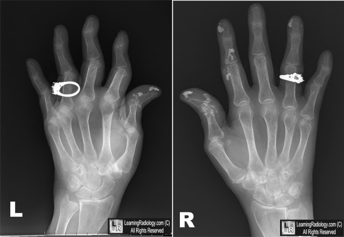
Calcinosis Circumscripta in Scleroderma. Radiographs of both hands show multiple, punctate calcifications (circles) in the soft tissues of both hands characteristic of calcinosis circumscripta. The patient had a 12 year history of scleroderma.
For this same photo without the arrows, click here
For more information, click on the link if you see this icon 
JH Klippel, LJ Crofford, JH Stone, CM Weyand. Primer on the Rheumatic Diseases12th Edition 2001; 353-367.
|
|
|
