|
|
Killian-Jamieson Diverticulum
General Considerations
- Arises from cervical esophagus below cricopharyngeal muscle (about C5-C6) laterally
- Originates on the antero-lateral wall of cervical esophagus through a muscular gap (the Killian-Jamieson space) lateral to longitudinal muscle of the esophagus
- It is a pulsion, not a true, diverticulum since it does not involve all layers of esophagus
- Usually smaller than a Zenker diverticulum
Clinical Findings
- Usually asymptomatic
- May produce reflux and aspiration
Imaging Findings
- Smooth-walled, contrast-containing pouch on barium swallow
- Most often unilateral and left-sided
- May be bilateral
Differential Diagnosis
- Zenker’s diverticulum
- Arises above cricopharyngeal muscle in midline posteriorly
- Develops at anatomically weak posterior zone (Killian’s dehiscence)
Treatment
- Only patients who are symptomatic or who have large diverticula are offered treatment
- Treatment options are either surgical or endoscopic

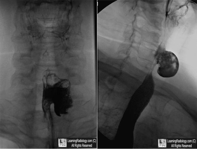
Killian-Jamieson Diverticulum. Left: Frontal view from a barium swallow shows an outpouching of barium (white arrow) arising laterally from the cervical esophagus. Right: The diverticulum (yellow arrow) is anterior to the normal esophagus.
For this same photo without the arrows, click here
For more information, click on the link if you see this icon 
Killian-Jamieson Diverticula: Radiographic Findings in 16 Patients. SE Rubesin and MS Levine. AJR July 2001 vol. 177 no. 1 85-89
Killian-Jamieson Diverticulum: The Rarer Cervical Esophageal Diverticulum. CH Chea, SL Siow, TW Khor and NA Nik Azim. Malaysia Vol 66 No 1 March 201
|
|
|
