|
|
Avascular Necrosis of the Scaphoid (Navicular)
General Considerations
- Scaphoid is the bone of the wrist most frequently fractured
- Most often is adults aged 15-60
- Some 10% of scaphoid fractures have associated fractures of other bones
- Most frequently radial styloid, but also triquetrum, capitate and perilunate fracture-dislocations
- About 70% involve the waist of the scaphoid, 10-20% the distal pole and 5-10% the proximal pole in adults
- In children, the tubercle is most often fractured
- Mechanism of injury is usually fall on an outstretched hand (FOOSH) but may include direct impact
- Complications include
- Malunion
- Most frequently angular deformity
- Delayed union
- Fracture still present after more than 4 months of immobilization
- Nonunion
- Fractures lines are smooth and sclerotic
- Avascular necrosis
- More common in scaphoid because of peculiar blood supply
- Up to 30% of scaphoid fractures may display increased density of the proximal pole
- Often reversible
- May be due to relative ischemia of proximal pole
- About 10% of scaphoid fractures display malunion or nonunion
- More common in fractures of the waist and proximal pole
- Delayed union occurs more frequently if fracture is not immobilized, especially after 4 weeks
- Treatment of delayed or nonunion
- About 90% of all acute scaphoid fractures heal if treated early
- Delayed union <6 months post-injury can be treated with prolonged immobilization and electrical stimulation
- Symptomatic nonunion is treated with bone graft
- Surgical removal of the proximal pole or whole scaphoid may be used
- Natural history of scaphoid nonunion is development of osteoarthritis
- May affect radiocarpal joint
- Scaphoid nonunion advanced collapse (SNAC), similar to scapholunate advanced collapse (SLAC) may occur with long-standing nonunion
- Vascular anatomy of the scaphoid
- Primary supply of blood comes from dorsal branch of radial artery which enters at the waist of the scaphoid
- Branches course proximally making the proximal pole at risk for AVN if the bloody supply is compromised at the waist or proximal pole by a fracture
- Occurs in 15-30% of scaphoid fractures
- Almost always involves proximal pole
- The more proximal is he fracture line, the risks of avascular necrosis increase
- The radiographic hallmarks of AVN are collapse and fragmentation
- MRI may be more sensitive to AVN than conventional radiographs but is not 100% sensitive
- Most common carpal instability pattern is scapholunate dissociation
- Scapholunate ligament may be disrupted and widening of the scapholunate space may occur
- Distance of 2-3mm is suggestive of disruption of the scapholunate ligament
- Widening of > 4mm is considered diagnostic of scapholunate ligament disruption


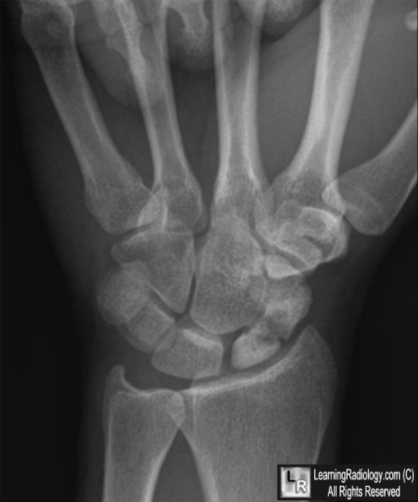
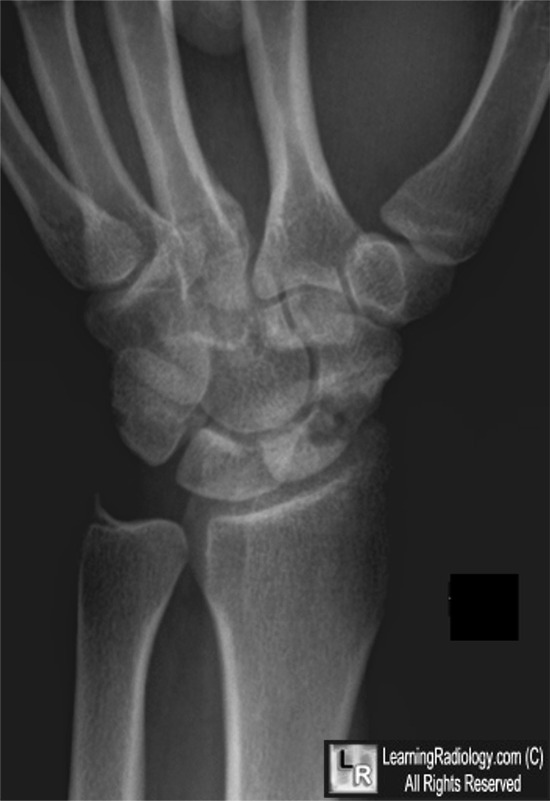
Avascular necrosis of the scaphoid secondary to fracture. Both photos: There is a transverse fracture of the waist of the scaphoid (yellow arrows) resulting in increased density of the proximal pole (white arrows) from avascular necrosis.
For these same photos without the arrows, click here and here
For more information, click on the link if you see this icon 
eMedicine Carol A Boles, MD Wrist, Scaphoid Fractures and Complications
|
|
|
