|
|
Pelvic Rib or Digit
General Considerations
- Rare congenital anomaly
- Bone develops in soft tissue
- Can arise from any of the pelvic bones; also anterior abdominal wall
- Most commonly arises from ilium
- Unilateral
- Believed to arise in mesenchymal stage of bone growth within first 6 weeks of embryogenesis
- May present as rib-like or finger-like structures with one or more (pseudo-) joints
Clinical Findings
- Usually asymptomatic; discovered incidentally
Imaging Findings
- Digit or rib has cortex and medulla
- “Finger” is typically segmented with pseudo-articulations
- May contain 2, 3 or 4 segments
- May or may not pseudo-articulate with the axial skeleton
Differential Diagnosis
- Heterotopic ossification – frequently a history of prior trauma
- Avulsion fracture of the pelvis
- Ossified scar – in subcutaneous tissue
- Fong's disease (onychoosteodysplasia) – iliac horns
Treatment


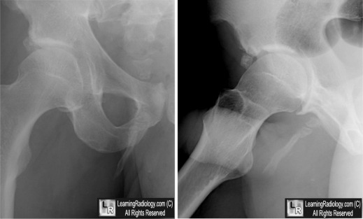
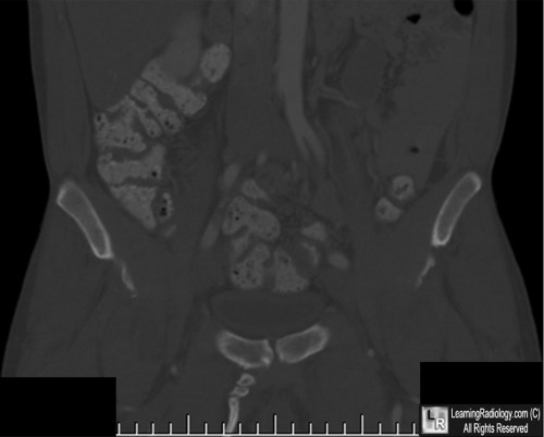
Pelvic Digit. (Above). Frontal and lateral radiographs of the hip demonstrate a well-corticated bony protuberance arising from the region of the right pubic bone with a suggestion of segmentation (yellow and white arrows). (Below) A coronal reformatted CT image of the pelvis confirms the presence of the pelvic digit with pseudo-articulations (white circle).
For these same photos without the arrows, click here and here
For more information, click on the link if you see this icon 
The pelvic digit: a harmless “eleventh” finger. IV Breuseghem. British Journal of Radiology (2006) 79, e106-e107
Pelvic digit as a rare cause of chronic hip pain and functional impairment: a case report and review of the literature. M Maegele. Journal of Medical Case Reports 2009, 3:139 doi:10.1186/1752-1947-3-13
|
|
|
