|
|
Cavitary Pulmonary Metastases
General Considerations
- Metastases to the lung occur in about 30% of all malignant disease
- Routes of spread include hematogenous, lymphangitic
and direct extension
- Most metastatic lung nodules develop through hematogenous spread
- Primary lung carcinomas cavitate more frequently than metastatic lesions to the lung
- Most cavitary metastases are epithelial in origin
- Squamous cell carcinomas are the most common metastases to cavitate (70%)
- Especially head and neck tumors
- Pulmonary lymphoma can rarely cavitate and almost always demonstrates associated adenopathy
Clinical Findings
- Can be asymptomatic, especially slow-growing malignancies, e.g. papillary thyroid cancer or adenoid cystic carcinoma of the salivary gland
- Later in course cancer is commonly dominated by signs and symptoms of advanced/terminal malignant disease and by signs and symptoms associated with primary tumor
Imaging Findings
- Chest radiographs are usually first examination to detect pulmonary metastases
- CT scanning has higher resolution than radiography, showing more and smaller nodules
- Plain radiographs detect cavitation in lung metastases in 4% of cases
- Metastases are multiple
- They are usually of differing sizes indicating different times of tumor embolization
- They usually have thick and irregular walls
- Thin-walled cavities may be seen with adenocarcinomas and sarcomas
- Cavitation is more common in upper lobe lesions than lower lobe
Differential Diagnosis
- TB
- Rheumatoid nodules
- Necrotizing granulomatosis
Treatment
- Chemotherapy is the usual treatment for disseminated tumor
- Pulmonary metastasis surgical removal can be performed depending on the underlying primary malignancy and surgical selection criteria
Prognosis
- Presence of pulmonary metastases is a poor prognostic indicator indicating disseminated disease
- Mortality depends on primary tumor


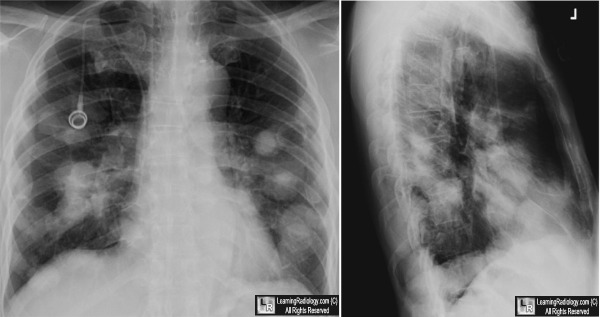
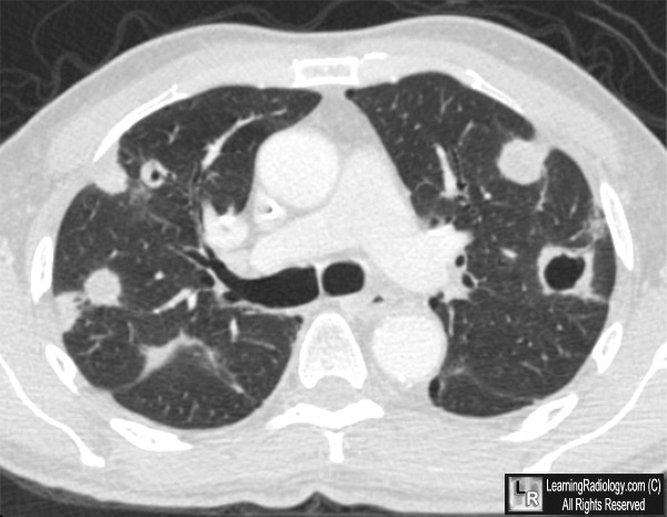
Cavitary Metastases to the Lung. Frontal and lateral chest radiograph (above) show multiple masses
in both lungs. At least one mass in the left lung (white arrow) is seen to be cavitary. An axial CT scan of the same patient demonstrates multiple masses, two of which show obvious cavitation (white arrows).
For these same photos without the arrows, click here and here
For more information, click on the link if you see this icon 
Cavities in the Lung in Oncology Patients. Medscape. RR Gill, S Matsusoka and H Hatabu.
Secondary Lung Tumors. Medscape. DS Schwartz and CS Chin.
|
|
|
