|
|
Retrocaval (Circumcaval) Ureter
General Considerations
- Also known as “circumcaval ureter”
- Abnormality in embryogenesis of IVC
- Results from abnormal persistence of right subcardinal vein positioned ventral to ureter in the definitive IVC
- Developing right ureter courses behind and medial to the IVC
- Incidence
- 0.07%
- Male to female ratio of 3:1
Clinical Findings
- Symptoms of right ureteral obstruction
Imaging Findings
- Normal course of ureters
- About the width of your thumb lateral to the lumbar vertebral pedicles
- About the width of two fingers medial to pelvic brim in true pelvis
- With retrocaval ureter
- Right ureter’s course swings medially over pedicle of L3/4
- Then exits anteriorly between IVC and aorta returning to its normal position in lower third
- Produces varying degrees of proximal hydroureteronephrosis
- Can be associated with Turner’s syndrome
- Types of retrocaval ureter
Types of Retrocaval Ureter |
Type 1 |
More common. Ureter crosses behind IVC at level of third lumbar vertebra; has fish hook shape at point of obstruction.
Marked hydronephrosis in 50% |
Type 2 |
Less common. Ureteral crossover higher at level of renal pelvis.
Mild hydronephrosis in majority of this type |
Differential Diagnosis
Causes of Ureteral Deviation or Displacement |
Medial Displacement or Deviation |
Upper ureter |
Lower ureter |
Retrocaval ureter |
Lymphadenopathy |
Retroperitoneal fibrosis |
Iliac artery aneurysm |
|
Bladder diverticulum |
|
Post-surgical (esp. AP resection) |
|
Pelvic lipomatosis |
Lateral Displacement or Deviation |
Upper ureter |
Lower ureter |
Lymphadenopathy |
Pelvic mass, e.g. uterine fibroids |
Aortic aneurysm |
|
Retroperitoneal hematoma |
|
Treatment
- Surgery may be done if there is hydronephrosis or recurrent infection


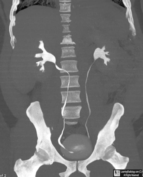
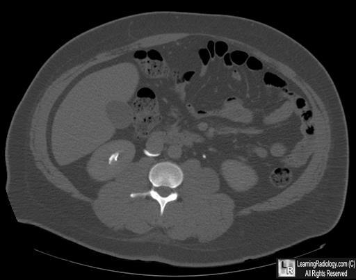
Retrocaval ureter. Coronal reformatted MIPS image from a CT urogram demonstrates medial
displacement of the right ureter at the level of L3 (White arrow). The ureter is medial to the pedicle
of the vertebral body (blue arrow). The Axial CT (below) shows the inferior ureter (white arrow)
coursing posterior to the inferior vena cava (red arrow).
For this same photo without the arrows, click here and here
For more information, click on the link if you see this icon 
Herman, T and McAlister, W: Radiologic Clinics of North America. Vol. 29:2, March, 1991.
Retrocaval ureter. B. B. Hyams, C. Schneiderman, and A. B. Mayman. Can Med Assoc J. 1968 January 6; 98(1): 45–49
|
|
|
