|
|
Arterial Placement of Central Venous Catheter
CVC, CVP
General Considerations
- Percutaneous cannulation of a central venous structure is a common procedure, most often for fluid replacement, administration of medication, for parenteral alimentation
- Arterial puncture has been reported in 5.2% of attempted central venous catheter (CVC) insertions
- Pneumothorax is the most common complication of central venous catheter insertion attempts
- Puncture of the artery (carotid) is more likely to occur in an attempted internal jugular approach than a subclavian vein approach for the subclavian artery
- Frequency of a mechanical complication increases six-fold when three or more attempts are made
- Arterial cannulation may occur if the subclavian puncture is too lateral or too deep
Clinical Findings
- Bright red pulsatile back flow instead of steady venous backflow
- Occurs more often with right-sided insertions
Imaging Findings
- Ultrasound-guided subclavian vein cannulation has reduced complications
- Chest radiography following attempted insertion of a CVC is the standard of care
- The normally-positioned CVC descends around the medial end of the clavicle
- The normal position of the tip of a CVC is to the right of midline at the first intercostal space
- Arterial placement is to be suspected if the catheter fails to descend to the right of the spine and/or…
- If the catheter follows the expected course of the aortic arch
Differential Diagnosis
- Persistent left superior vena cava
- Inadvertent arterial puncture and a right-sided aortic arch
Treatment
- Most can be managed percutaneously
- For subclavian arterial punctures, patients need to be monitored for hemodynamic instability and hemothorax development
Complications


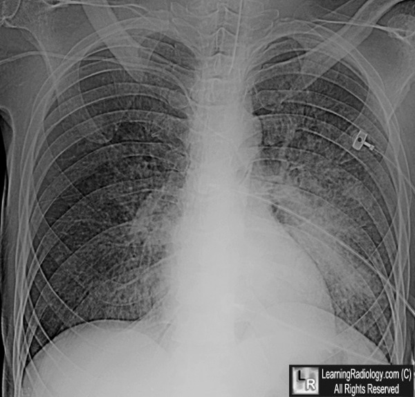
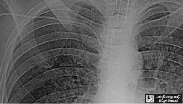
Arterial Placement of a central Venous Catheter. A percutaneously inserted line is seen to curve
downward at the medial end of the clavicle (C) but then cross the midline and descend on
the left side of the spine (white arrows). The anterior, medial end of the first rib is labeled 1.
For these same photos without the arrows, click here and here
For more information, click on the link if you see this icon 
Complications of 1303 central venous cannulations. A Yilmazlar, H Bilgin, G Korfali, A Eren, and U Ozkan. J R Soc Med. 1997 June; 90(6): 319–321.
Central Venous Catheterization: Concise Definitive Review. RW Taylor; AV Palagiri.
Crit Care Med. 2007;35(5):1390-1396. Lippincott Williams & Wilkins.
Complications of central venous catheterization. SE Mitchell and RA Clark. American Journal of Roentgenology, Vol 133, Issue 3, 467-476
|
|
|
