|
|
Neurocysticercosis
General Considerations
- Most common parasitic infestation of central nervous system worldwide
- Caused by the pork tapeworm, Taenia solium
- Most important manifestation of disease is in central nervous system-neurocysticercosis (NCC)
- Endemic areas include Mexico and Latin America, sub-Saharan Africa, India and east Asia
- Incidence in USA is increasing from immigration and travel
- Life cycle
- Humans are definitive host of intestinal tapeworm
- Pigs are intermediate host
- Human cysticercosis develops when eggs or larva are ingested through fecal-oral transmission from a tapeworm host or in undercooked pork
- Cysticerci (larval cysts) develop within organs
- Skin, skeletal muscle, CNS, heart, eye

Life Cycle of Taenia Solium (Pork Tapeworm) (From CDC)
Clinical Findings
- Dependent on pericystic inflammation
- If no inflammation, usually asymptomatic
- Seizures
- Chronic headache
- Nausea and vomiting
- Mental status change
- Vision changes
Imaging Findings
- Usually parenchymal in brain
- May be ventricular or subarachnoid
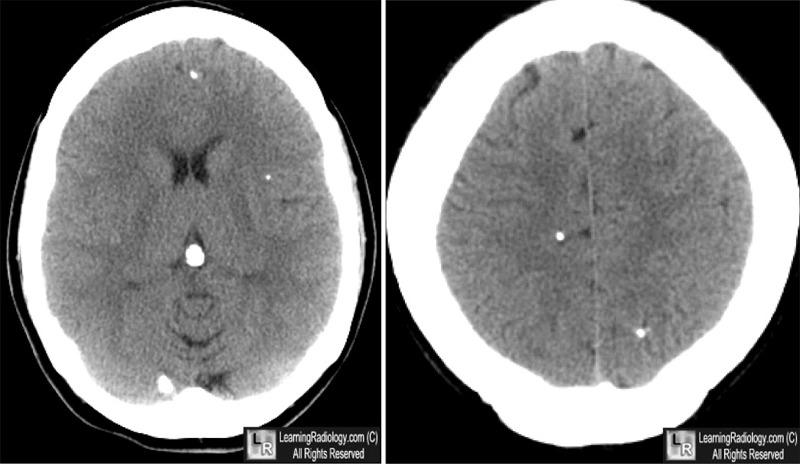
- Involuted, degenerated cysts calcify and appear oblong or cigar-shaped
- CT is the imaging study of choice for NCC
- Calcified cysts are usually not associated with surrounding inflammation
- Non-enhancing cystic lesions with/without surrounding edema
- Mass effect
- Hydrocephalus
- MRI may demonstrate cysts with then ventricular system
- Lateral, 3rd and 4th ventricles
Differential Diagnosis
- Coccidiomycosis
- Toxoplasmosis
- Tumor
- Trichinosis
- TB
Treatment
- Anti-helminthic medications
Complications
- Encephalopathy
- Obstructive hydrocephalus
- Sequelae of treatment from steroids or anti-convulsants
- Calcified cysts may be epileptogenic
Prognosis
 
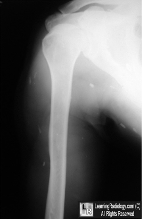
Cysticercosis. White arrows point to calcified cysticerci scattered throughout the
brain parenchyma with no surrounding inflammation. The right shoulder demonstrates
multiple cigar-shaped, muscular calcifications, again representing cysticerci.
For more information, click on the link if you see this icon 
For these same photos without the annotations, click here and here
Cysticercosis: Differential Diagnoses & Workup. eMedicine. Ryan Tenzer, R and Blumstein, H.
|
|
|
