|
|
Lipomatous Hypertrophy of the Interatrial Septum
General Considerations
- Less rare than originally thought
- Benign accumulation of unencapsulated collection of brown fat in interatrial septum
- Hyperplasia more than hypertrophy of fat cells
- Associated with older age and obesity
Clinical Findings
- Asymptomatic in most
- Atrial arrhythmias
Imaging Findings
- Imaging modalities include transthoracic or transesophageal echo, CT or MRI
- The latter allow for tissue characterization and are more accurate in assessing extent of lesion
- CT
- Non-enhancing, dumbbell-shaped mass of fat density
- Confined to interatrial septum
- Spares fossa ovalis producing dumbbell shape
- Cephalad portion thicker than caudal portion of mass
- MRI
- Homogenous, bilobed high signal mass
- Associated with mediastinal lipomatosis
Differential Diagnosis
- Myxoma
- Rhabdomyoma
- Fibroma
- Lipomas
- Very rare, well-encapsulated but may be the same entity as lipomatous hypertrophy
Treatment
- Surgical resection has been performed but is usually not required
Complications
- Can cause right atrial obstruction
- Supraventricular arrhythmias
- Unexpected cardiac death
 
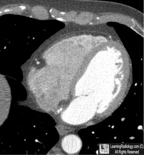
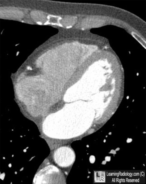
Lipomatous Hypertrophy of the Interatrial Septum. Axial, contrast-enhanced, CT scans
through the heart demonstrate a fatty mass in the interatrial septum (blue and red arrows) which has a dumbbell shape to it (blue arrow). sparing the fossa ovalis.
For more information, click on the link if you see this icon 
For these same photos without the annotations, click here and here
Lipomatous Hypertrophy of the Interatrial Septum: An Overview. O'Connor, S; Recavarren, R; Nichols, L and Parwani, A. Archives of Pathology & Laboratory Medicine. March 2006
CT appearance of lipomatous hypertrophy of the interatrial septum. Meaney, J; Kazerooni, E; Jamadar D and Korobkin, M. American Journal of Roentgenology, Vol 168, 1081-108
|
|
|
