|
|
Typhlitis
Neutropenic Enterocolitis
Submitted by Zombor Zoltani, MSIV
General Considerations
- Seen in neutropenic patients undergoing immunosuppressive therapy for malignancy
- May also present in patients with aplastic anemia, AIDS, lymphoma, or after immunosuppressive therapy s/p solid organ transplant
- True incidence is unknown
-
Pathogenesis
- Mechanism of disease not well understood
- Implicated factors are mucosal injury, neutropenia, impaired immune response to pathogen infiltration leading to necrosis of bowel wall
- Cecum thought to be uniquely affected due to ease of distensibility, relative stasis of bowel contents, and decreased vascularity relative to remainder of colon
Clinical Findings
- Fever, watery/bloody diarrhea, right lower quadrant abdominal pain
- Initial symptoms often occur 10-14 days following cytotoxic therapy
- Suspect in patient with severe neutropenia (absolute neutrophil count < 500)
Imaging Findings
- CT is study of choice
- Cecal distension and circumferential thickening of cecal wall
- Mesenteric fat stranding secondary to inflammation
- Submucosal bowel wall edema
- Paralytic ileus
- Terminal ileum and ascending colon may also be affected
- Pneumatosis, pneumoperitoneum, and pericolic fluid may be seen and indicate urgent need for surgical intervention
- Abdominal Radiograph
- Right lower abdominal mass density
- Paralytic ileus or obstruction
- Barium Enema
- In general, avoid barium enema or colonoscopy due to risk of perforation
- Thick walled and poorly distensible cecum
- Thumbprinting
Differential Diagnosis
Treatment
- Uncomplicated disease
- Treated with bowel rest, total parenteral nutrition, IV fluids, and broad spectrum antibiotics
- In cases of peritonitis, perforation, or GI bleeding
- Surgical management with right hemicolectomy
- CT may be used to follow treatment
- Bowel wall thickness will decrease
Prognosis
- Mortality rate is 40-50%
- Cause of death linked to transmural bowel necrosis, bowel wall perforation, and sepsis


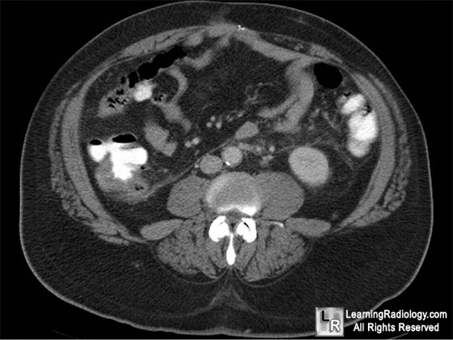
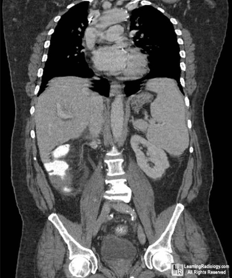
Typhlitis. Axial and coronal CT images of the lower abdomen demonstrate
thickening of the wall of the cecum, nodularity of the lumen and pericolonic stranding
consistent with typhlitis in this immunocompromised individual.
For more information, click on the link if you see this icon 
For this same photo without the annotations, click here and here
Horton KM, Corl FM, and Fishman EK. CT Evaluation of the Colon: Inflammatory Disease. Radiographics. March 2000. 20:399-418.
Song LM and Marcon NE. Necrotizing enterocolitis (typhlitis) in adults. In: UpToDate. Basow, DS (Ed), UpToDate. Waltham, MA. 2009.
Frick MP, Maile CW, Crass JR et al. Computed Tomography of Neutropenic Colitis. American Journal of Roentgenology. October 1984. 143:763-765
|
|
|
