|
|
Accordion Sign
General Considerations
- The sign was originally described as alternating edematous haustral folds separated by mucosal ridges filled with oral contrast material
- Simulated appearance of an accordion
- It was first thought to be specific for severe Clostridium difficile–related colitis (pseudomembranous colitis)
- Almost all cases of pseudomembranous colitis are associated with recent antibiotic therapy
- C. difficile is the largest single cause of this sign
Clinical Findings
- C. difficile is a gram-positive anaerobic bacillus
- Can cause a spectrum of GI diseases ranging from mild diarrhea to fulminant life-threatening colitis
Imaging Findings
- The sign is due to the marked degree of colonic wall thickening caused by the pseudomembranes and edematous tissues that develop in C difficile colitis
Differential Diagnosis
- Cirrhosis with colonic edema
- Ischemic colitis

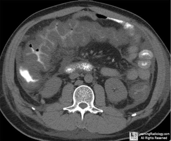
Accordion Sign. White oval highlights markedly thickened bowel wall
with oral contrast
trapped between haustral folds in a patient with known C difficile colitis. This is the "accordion sign."
For more information, click on the link if you see this icon 
For this same photo without the annotations, click here
The Accordion Sign at CT: a Nonspecific Finding in Patients with Colonic Edema Macari, M; Balthazar, E and Megibow, A. Radiology June 1999 211:743-746
|
|
|
