|
|
Meckel’s Diverticulum
Submitted by Denise Drohobyczer MS IV
General Considerations “Rule of 2”
- Most common congenital anomaly of GI tract
- Within 2 feet proximal to ileocecal valve
- 2 inches long, 2 cm wide
- 2 types of heterotopic mucosa
- Symptomatic in 2%
- M>F (2:1)
- Children > adults; often < 2 years old
Embryology and Pathology
- Incomplete atrophy of omphalomesenteric (vitelline) duct
- True diverticulum on anti-mesenteric border of ileum
- 50% contain heterotopic mucosa
- Most often gastric (50%) and pancreatic (16%)
Complications and Clinical Findings
- Hematochezia (~40% of complications)
- Most common complication in children
- Heterotopic gastric mucosa ® ulceration and hemorrhage
- Occult blood loss and iron deficiency anemia (adults)
- Small bowel obstruction (~35%)
- Most common complication in adults
- Abdominal guarding and distension, vomiting, obstipation
- Causes: Volvulus (with persistent fibrous band), torsion, foreign body, Littre’s Hernia (inclusion in hernia sac), inversion ® intussusception
- Diverticulitis (~17%)
- Second most common complication in adults
- RLQ or periumbilical acute abdominal pain
- Can result in perforation
- Enteroliths (~10%)
- Neoplasia (~3%)
- Most often carcinoid (M>F)
Imaging Findings
- Abdominal Film, non-specific:
- Enteroliths, small bowel obstruction, perforation
- Abdominal CT
- Diverticulum off distal ileum, small bowel obstruction, intussusception, perforation, diverticulitis, necrosis, enteroliths or inversion of tic
- Sonography
- Obstruction, inversion and intussusception
- Enteroclysis
- “Triradiate” fold pattern
- Junction of omphalomesenteric duct and ileum
- Rugal pattern like stomach(heterotopic gastric tissue)
- Meckel’s scan (Technetium-99m pertechnetate scintigraphy)
- Gastric mucosa absorbs and secretes radioactive isotope
- “Hot spot” in RLQ- most commonly
- Gold standard for hemorrhagic diverticula
- 95% specific in children, 60% specific in adults
- Angiography
- Visualization of remnant omphalomesenteric (vitellointestinal) artery
- Off distal ileal branch of superior mesenteric artery
- Vascular blush (ischemia)
Differential Diagnosis
- Appendicitis, colonic diverticulitis, acute mesenteric lymphadenitis
- Single small intestinal diverticulum
- Communicating enteric duplication
- Pseudosacculations (Crohn’s Disease)
- Cavitating malignancies (lymphoma, GIST)
Treatment
- Surgical excision if symptomatic
- Prophylactic removal is controversial

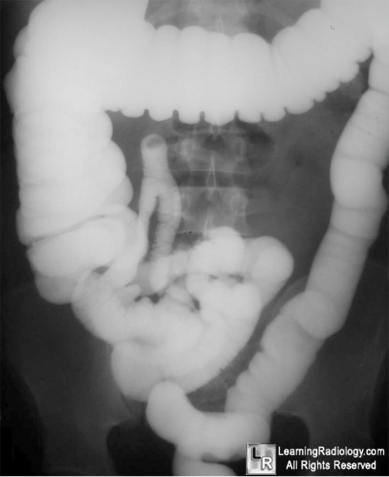
Meckel's Diverticulum. Reflux into the small bowel has occurred during a single-contrast barium enema examination. Black arrow points to Meckel's diverticulum arising from small bowel near terminal ileum.
For more information, click on the link if you see this icon 
For this same photo without the annotations, click here
Levy AD, Hobbs, CM. Meckel Diverticulum: Radiologic Features with Pathologic Correlation. Radiographics. 2004; 24: 565-587.
O’Neill, J, Grosfeld, J. Principles of Pediatric Surgery. Mosby, 2003.
|
|
|
