|
|
Hydatid Cyst of the Liver
Cystic Echinococcosis
General Considerations
- Parasitic infestation caused by Echinococcus tapeworm
- Endemic to Middle East, Africa, Iceland, Australia, southern parts of South America
- Cysts grow slowly and usually don’t become symptomatic until adulthood (30-50 years old)
- Three species which cause human disease are
- E. granulosus (aka granulosa, granulosis) causes Cystic Echinococcosis (CE) (most common)
- E. multilocularis causes Alveolar Echinococcosis (AE) –most virulent
- E. vogeli—rarest
- Infestation of humans, who are accidental intermediate hosts, occurs from ingestion of water or food contaminated by fecal material of definitive hosts (dogs, wolves, deer, sheep, coyotes)
- Eggs hatch in duodenum and pass as larvae through intestinal wall into blood stream to liver and lungs
Clinical Findings
- Asymptomatic in many cases
- Most symptomatic cysts are 5 cm or more
- Symptoms frequently come from pressure on affected organs
- Jaundice, abdominal pain if in liver
- Cough, dyspnea and chest pain if in lungs
- Other symptoms arise from leakage of cyst contents or infection of cyst
- Pain, flushing and urticaria
- Anaphylactic reaction
- Fever and sepsis
- Liver is most common organ involved (mostly right lobe)
- Then, lungs, muscles, bones kidneys, brain and spleen, in order
Imaging Findings
- Conventional radiographs
- May show a rim-like calcification in liver cyst
- CT is most accurate
- Low density, fluid-filled masses
- May have septations
- Wall may be calcified or enhance with contrast
- Wall is frequently of high density even without contrast-enhancement
- Internal calcifications may also occur
- There may be multiple daughter cysts
- Inner layer of cyst may separate from outer layer and appear as a serpentine structure inside of the cyst
- Water-lily sign is characteristic of hydatid cyst
- Multilocular cysts may have a honeycomb pattern from septations
- US
- Usually echogenic because of daughter cysts or infolded membranes
- Daughter cysts can be either echogenic or non-echogenic
- Fragments may be seen in common bile duct
Differential Diagnosis
Treatment
- For Cystic Echinococcosis, surgery is most frequently used
- Repeated aspiration of cysts with injection of anti-helminthic agent
- Chemotherapy includes use of benzimidazoles
Complications
- “Metastases” can occur in lung, brain or bone
- Complications, including anaphylaxis, come from
- Rupture of cyst (intra-biliary rupture is most common)
- Infection of cyst
- Biliary obstruction
- Cirrhosis Bronchial obstruction
- Urinary tract obstruction
Prognosis
- With Cystic Echinococcosis, prognosis is good with surgical excision without spillage
- In Alveolar Echinococcosis, 10-year mortality rates of 10%


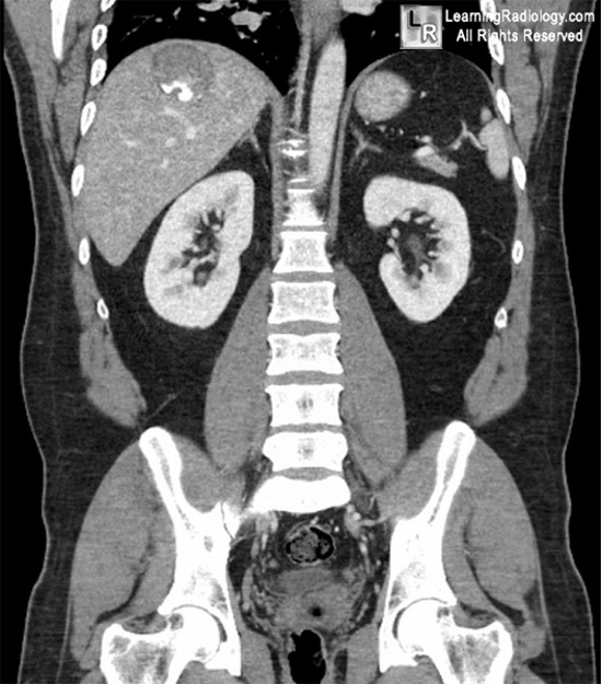
Hydatid Cyst of the Liver. Black arrows point to a cystic lesion on the right lobe of the liver containing dense calcifications. The cyst was excised.
For more information, click on the link if you see this icon 
For this same photos without the annotations, click here and here
Hydatid Cysts eMedicine Dandan, I; Soweid, A and Abiad, F
Imaging Appearances Of Hydatid Cyst Ds Shah, H Parikh, B Shah, S Banuprakash, J Shah Ind J Radiol Imag 2006 16:4:533-535
Hydatid cyst of the liver: rupture into the biliary tree. L Marti-Bonmati, F Menor, and A Ballesta. American Journal of Roentgenology, Vol 150, Issue 5, 1051-1053
|
|
|
