|
|
Intraosseous Pneumatocysts
General Considerations
- Almost always found in the ilium or sacrum, near the sacroiliac joint
- Also in the spine, especially cervical spine associated with degenerative disease
- Gas-containing
- The gas is mostly nitrogen
- Uncertain etiology
- A communication with the joint is usually not visible
Imaging Findings
- They are rarely visible on conventional radiographs
- CT will identify the lesions as containing gas
- Lesions have a sclerotic margin
- Low density on CT scans
- Decreased attenuation on both T1 and T2-weighted images
- May be confusing on MR alone as they can be indistinguishable from blastic metastases
Differential Diagnosis
- Pathognomonic on CT
- Osteomyelitis produced by a gas-forming organism
- Osteonecrosis
Complications
- Some pneumatocysts in the spine have been shown to enlarge
- Other have been shown over serial studies to fill in with fluid

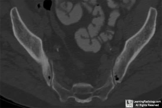
Pneumatocysts. The white arrows point to two air-containing cyst-like structures
in the ilii adjacent to the sacroiliac joints on an axial CT image of the pelvis. The lesions have
a characteristic sclerotic margin.
For more information, click on the link if you see this icon 
For this same photo without the annotations, click here
Vertebral pneumatocysts: uncommon lesions with pathognomonic imaging characteristics. Haithcock, J, with Layton, K andOpatowsky, M. Proc (Bayl Univ Med Cent). 2006 October; 19(4): 423–424.
Natural Course of an Intraosseous Pneumatocyst of the Cervical Spine. Yamamoto, T; Yoshiya1, S; Kurosaka1, M; Nagira, K; Takabatake, M; Hamamoto, H and Kazuo Mineo, K. AJR 2002; 179:667-669
Enlarging Vertebral Body Pneumatocysts in the Cervical Spine. Kitagawaa, T; Fujiwaraa, A; Tamaia, K; Kobayashia, N; Saikia, K; Omataa, S and Saotomea, K. American Journal of Neuroradiology 24:1707-1710, September 2003
|
|
|
