|
|
Melorheostosis
Leri’s Disease, Flowing Periosteal Hyperostosis
General Considerations
- One of a group of sclerosing bone disorders
- Rare
- Cause is unknown
- Produces thickening of the endosteum and periosteum
- Peak age of presentation is 5-20 years
- May be monostotic or polyostotic
- May involve one entire limb
- Usually does not involve multiple limbs
- Twice as common in lower extremities than elsewhere
Clinical Findings
- About 50% affected develop symptoms by age 20
- Adults present with
- Joint stiffness
- Deformity that may progress over time
- Children may present with
Imaging Findings
- Patterns described include:
- Candle-wax appearance (classic)
- Resembling myositis ossificans
- Resembling osteopathia striata
- Mixed
- Sclerotic lesions of cortical bones, usually in the diaphysis, that resemble “candle-wax-dripping”
- Cortical hyperostosis with an undulating appearance usually affecting one side of a bone
- Soft tissue lesions that may calcify
- Adjacent to involved bone
- May grow to compress nerves
- Usually low signal on MRI
- Bone scan is markedly positive
Differential Diagnosis
- Osteopathia striata
- Longitudinal dense striations
- Osteopoikilosis
- Punctate, rounded bone islands surrounding joints
- Osteosarcoma
Treatment
- Analgesia
- Nerve blocks
- Surgery directed at relieving contracture
Complications
- Associated soft tissue lesions and cutaneous lesions
- Vascular malformations
- Neurofibromatosis
- Tuberous sclerosis
- Hemangioma
- Muscle contractures
- Scoliosis
Prognosis
 
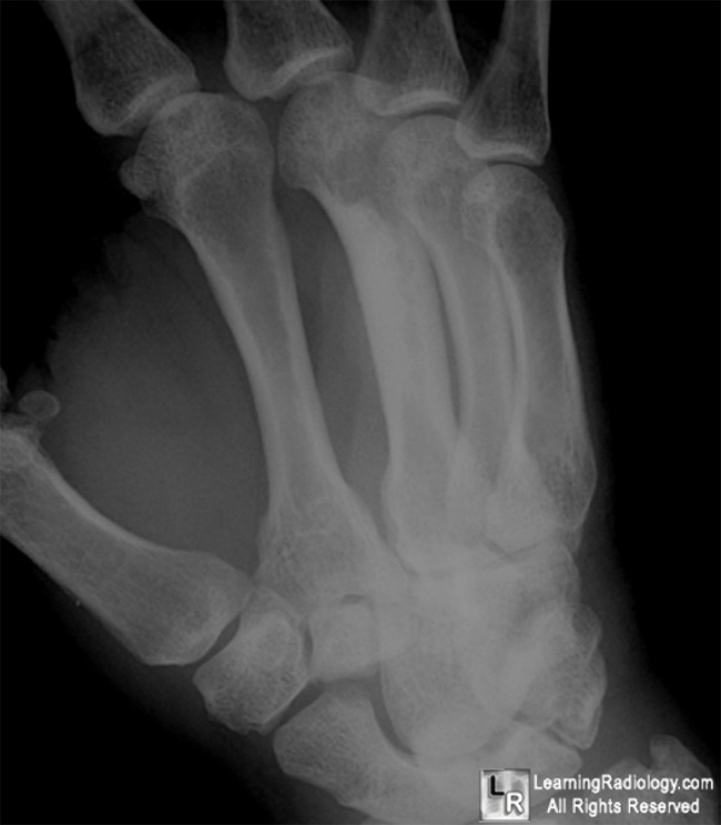
Melorheostosis. Frontal and oblique radiographs of the hand shown in close-up demonstrate the
undulating cortical hyperostosis representing the classical "candle-wax" dripping appearance
involving only the radial side of the 3rd metacarpal shaft.
For more information, click on the link if you see this icon 
For this same photo without the annotations, click here and here
BoneTumor.org DeGroot, H.
|
|
|
