|
|
Carcinoid Tumor of the Small Bowel
Submitted by Theresa Kaufman, MSIV
General Considerations
- Rare, potentially malignant, neuroendocrine tumor of primitive stem cells in gut wall which have hormone-secreting potential
- Arise from enterochromaffin cells of Kulchitsky
- Male predominance
- ~50% of carcinoids occur in appendix; ~33% occur in small bowel
- Most common primary tumor of the small bowel
- Most appendiceal carcinoids are benign
Clinical Findings
- Most are asymptomatic in early development
- Most common presentation is episodic abdominal pain
- Secretory products:
- Serotonin
- Histamine
- Kallikrein
- Prostaglandins
- Serotonin is deaminated by monoamine oxidase in liver and lungs to 5-hydroxyindoleacetic acid (5-HIAA)à excreted in urine
- Normal: <10 mg 5-HIAA in 24-hour urine
- Serotonin mediates desmoplastic change, leading to fibrosis
- Metastases to mesenteric lymph nodesàalso produce endocrine substances and fibrosis
- Fibrosis is characteristically “spoke-wheel” toward adjacent bowel loops, causing them to pull closer
- Carcinoid syndrome
- Usually associated with liver metastasis (see Complications below)
Imaging Findings
- Usually performed once biochemical diagnosis confirmed, typically by elevated 24-hour excretion of 5-HIAA
- Barium studies are non-specific and less helpful than CT and scintigraphy
- Smooth intraluminal, rounded, asymmetric mass, usually in ileum
- Tethering of bowel loops by fibrosis, causing bending of bowel wall and crowding of folds
- Abdominal CT with intravenous and oral contrast
- Visualization of primary tumor
- Lymph node enlargement—mesenteric, para-aortic or retroperitoneal
- Radiating linear strands around soft tissue mass due to fibrosis (“spoke-wheel” pattern); may contain calcification
- Bowel wall thickening of adjacent bowel loops
- Liver metastases

- Often multiple
- May be hypervascular
- May become calcified
- Frequently associated with carcinoid syndrome
- 2% of tumors <1 cm in diameter metastasize; 80% of tumors >2 cm metastasize
- Ischemia or obstruction due to fibrosis may be present
- Indium-111 Octreotide imaging (Octreoscan)
- Octreotide is a somatostatin analogue
- Carcinoid tumor cells almost always contain somatostatin receptors, and show increased uptake on scan
- More sensitive than MIBG scans
- MRI with gadolinium
- Useful for detection of metastases
- Low T1 and high T2
- Enhance peripherally in arterial phase and hypointense during portal venous phase
Treatment
- Surgical resection with mesenteric lymph node excision
- For all tumors, even with metastasis, to prevent development/ progression of fibrosis and other complications
- Management of symptoms
- Patients with severe flushing and/or diarrhea à somatostatin analogue, octreotide
- Symptoms resistant to octreotide aloneà add IFNa
Complications
- Carcinoid syndrome
- Usually occurs with liver metastasis
- Vasoactive substances released into systemic venous flow
- Signs and symptoms include
- Cutaneous flushing (early and frequent sign with metastases)
- Secretory diarrhea (84%)
- Bronchospasm/asthma (25%)
- Carcinoid crisis
- Severe flushing, changes in blood pressure, bronchoconstriction, arrhythmias, confusion/stupor
- Carcinoid heart disease
- Right-sided subendothelial fibrosis
- Tricuspid stenosis/insufficiency
- Pulmonic stenosis
- Left side of heart is protected by monoamine oxidase in lungs
- Intussusception
- Carcinoid may be the lead point
- Small bowel obstruction
- Small bowel ischemia
- Has an association with scleroderma
Prognosis
- Dependent upon site of origin, size, presence/extent of metastases, histology, presence of carcinoid syndrome
- Complete resection of small bowel carcinoid à 75% 5-year survival rate
- Lower (19%) survival rate in patients with distant metastases


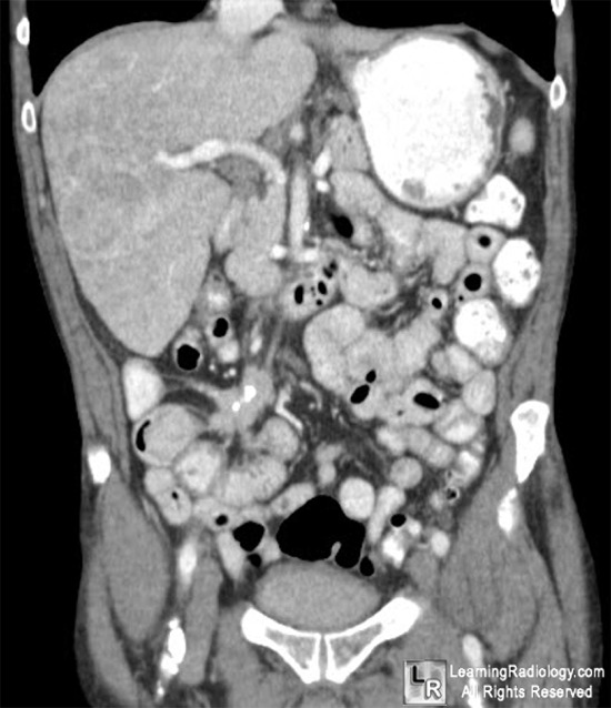
Carcinoid tumor of appendix. The white arrows are pointing to a soft tissue mass in the right lower
quadrant containing calcium and surrounded by fibrotic stranding (blue arrows).
For more information, click on the link if you see this icon
For this same photo without the annotations, click here and here
eMedicine Carcinoid Tumor Tebbi, C
|
|
|
