|
|
Benign Cortical Defect
Fibrous Cortical Defect, Non-ossifying fibroma
General Considerations
- Also called a non-ossifying fibroma or fibrous cortical defect
- Non-ossifying fibroma frequently reserved for lesions > 2cm in size in older children
- Usually arises in metaphysis of distal femur or tibia
- Solitary lesion (75%) or multiple (25%)
- Most commonly seen in children 2-15 years of age
- May be secondary to a prior trauma injury (traction) as they tend to occur at sites of insertion of tendons and ligaments
Clinical Findings
- Usually asymptomatic
- Found serendipitously
Imaging Findings
- Geographic lytic lesion
- Septated
- Metaphyseal
- Eccentric
- Well-marginated
- Sclerotic rim
- Endosteal scalloping
- On healing
- Marginal sclerosis increases
- Lesions “fill-in” from diaphyseal side of bone
- Bone scan
- May show increased activity on healing of the lesion
- MRI
- Variable signal intensity depending on healing stage
- Central decreased T2-W signal
- From collagen and hemosiderin deposits
Differential Diagnosis
- Image on conventional radiography is usually diagnostic
Treatment
Complications
- Rarely may undergo pathologic fracture
- Do not undergo malignant transformation
Prognosis
- Migrate away from epiphysis towards diaphysis with age
- Most lesions heal spontaneously by being replaced with normal bone


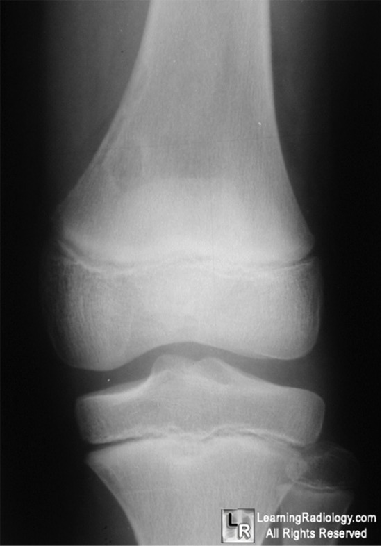
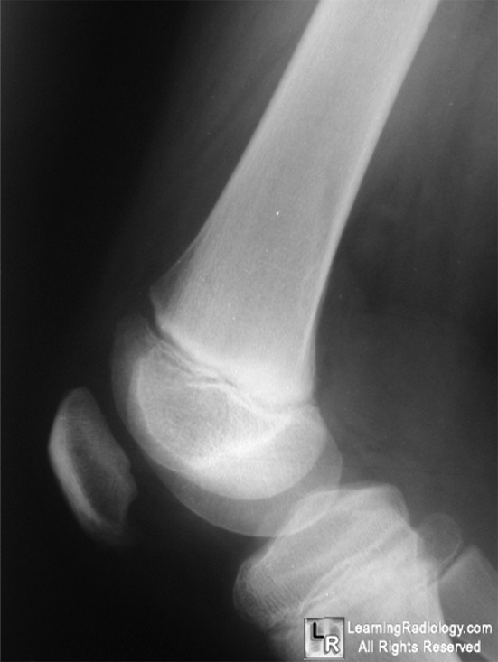
Benign Cortical Defect. White arrow (top) points to a well-circumscribed, lytic lesion in the metaphysis of this child's distal femur. It has a sclerotic rim. On the lateral view, its cortical nature is again demonstrated (yellow arrow).
For more information, click on the link if you see this icon 
For this same photo without the annotations, click here and here
|
|
|
