|
|
Abdominal and Pelvic Hernias
Inguinal, Femoral, Umbilical, Paraumbilical, Richter, Incisional, Spigelian and Obturator Hernias
Hernia Central

General Considerations
- By definition: protrusion of abdominal structures through the abdominal wall containing (1) an opening in the abdominal wall, and (2) a hernia sac consisting of abdominal contents enclosed by peritoneum
- Majority of abdominal hernias in adults that are acquired are iatrogenic (surgery)
- Inguinal hernias are most common, mostly indirect type
- High male to female ratio for inguinal hernias
Types
Groin (pelvic) hernias
Indirect inguinal hernia. There is a large inguinal (scrotal) hernia (white arrow) containing a dilated loop of small bowel (red arrow).
- Direct inguinal hernia
- Acquired defect in transversalis fascia of Hesselbach triangle
- Medial to inferior epigastric vessels
- More common in middle-aged males
- Femoral hernia
- Through the femoral canal medial to the femoral vein
- Only 4% of groin hernias (versus 96% inguinal)
- More often in females but still less common than inguinal hernias
- Right side more often than left
- Frequently incarcerated
Ventral (abdominal wall) hernias
- Umbilical hernia
- Failure of closure of the umbilical ring in children
- More females than males in adults
- More common in African Americans
- Incarceration is rare in children, more common in adults
- After infancy, second spike in incidence is in middle-age and result of
- Increased intra-abdominal pressure
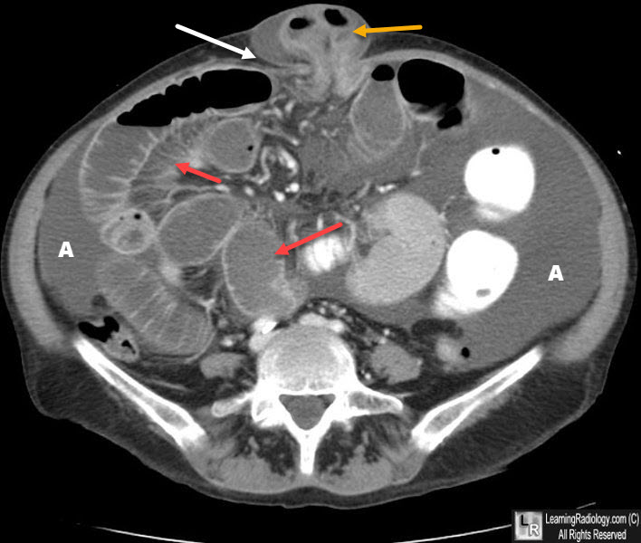
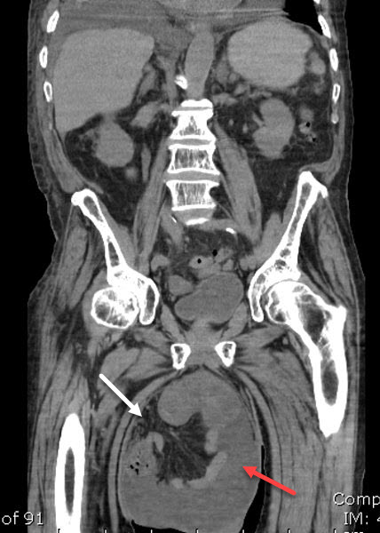
Umbilical Hernia. Mesenteric fat (white arrow) and bowel (orange arrow) protrude through the abdominal wall at the umbilicus. The hernia has caused a small bowel obstruction with dilated loops of small bowel seen (red arrows) There is also ascites present (A).
- Paraumbilical hernia
- Adults
- Occurs adjacent to site of umbilicus
- Superior to umbilicus called epigastric (more common)
- Inferior to umbilicus called hypogastric
- Occur along the linea alba
- Usually contain fat
- Richter hernia
- Involves only anti-mesenteric side of bowel entering hernia
- Usually no obstructive symptoms
- Can occur with any of the abdominal hernias
- Incisional hernia
- Breakdown in fascia closing prior abdominal surgery
- More common with obesity, wound infection and smokers
- Usually occur within first few months after surgery
- Can be quite large and are frequently incarcerated
- Spigelian hernia
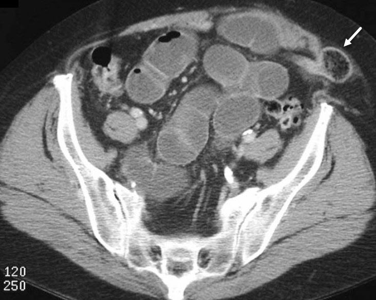
Spigelian Hernia. A loop of small bowel has herniated through the lateral edge of the rectus muscle at the semilunar line (white arrow) and produced a mechanical small bowel obstruction.
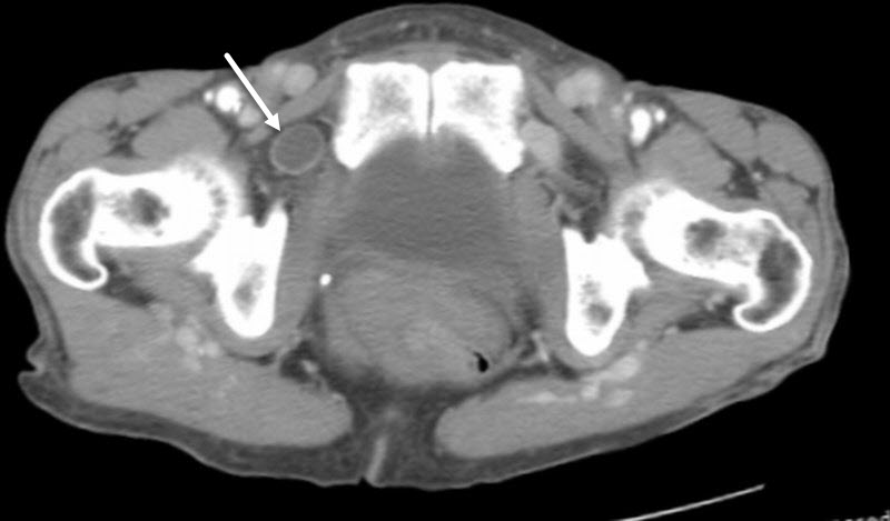
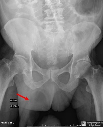
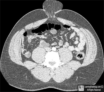
Obturator hernia. A loop of small bowel has herniated between the pectineus and obturator muscles (white arrow).
Clinical Findings
- Fullness at site of hernia
- Symptoms of bowel obstruction
- Symptoms of bowel ischemia
Imaging Findings
- Bowel loops projecting into scrotum or over obturator foramen on conventional radiographs

- Bowel and/or omentum (fat) or sometimes visceral organs protruding through peritoneum
- Bowel or fat in hernia sac
- Stranding of fat suggest the possibility of incarceration
- Proximal bowel dilatation from obstruction
- Bowel wall thickening, extraluminal fluid, severe fat stranding and engorged mesenteric vessels suggest strangulation
Differential Diagnosis
- Soft tissue tumors
- Lymphadenopathy
Treatment
- Surgery for incarceration
- Inguinal hernias are usually repaired to prevent incarceration and obstruction
- Spigelian and obturator hernias are usually repaired before they become symptomatic
Complications
- Obstruction
- Ischemic and/or necrotic bowel
- Perforation

Ventral (umbilical) hernia. Almost completely well-circumscribed, soft tissue mass (blue arrows) overlies umbilicus demonstrating incomplete rim sign (white arrow) where hernia is attached to body. Hernia contains fat, but appears dense because it is an additive density superimposed on the normal soft tissues of the abdomen.
For more information, click on the link if you see this icon 
For this same photo without the annotations, click here
|
|
|
