|
|
Skeletal Sarcoid
Osseous Manifestations of Sarcoidosis
General Considerations
- Radiographic evidence of osseous involvement occurs in about 5% of patients with sarcoidosis
- Arthralgia in patients with sarcoid may occur in up to 1/3 of patients
- Patients are usually between 20-40, more frequently women
- Typically affects multiple joints
- Most often affects hands, feet and ankles (the latter in acute form)
- Lesions in long bones, skull or spine are rare
- Often occurs in association with pre-existing pulmonary and/or skin lesions such as lupus pernio
- Purplish, indurated macules or papules on the face considered classic for sarcoid
- May occasionally be presenting manifestation of sarcoid

Clinical Findings
- Frequently asymptomatic
- Joint manifestations include
- Pain
- Soft tissue swelling
- Soft tissue nodules (granulomas)
- Erythema nodosum, especially in acute form
Imaging Findings
- Usually bilateral and symmetric
- Middle and distal phalanges of hands and feet most frequent sites of involvement
- Tends to spare the wrists
- Distal portions of small bones of hands and feet tend to be affected first
- Patterns of osseous sarcoidosis
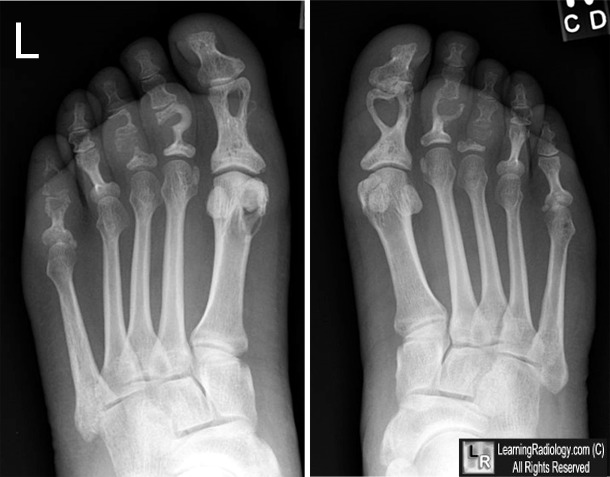
- Small, cortical, punched-out lytic lesions, usually well-corticated, in the phalangeal heads are most common manifestation
- Some been described as heart-shaped as in this case
- Reticular (permeative) pattern produces a lace-like (driftwood) pattern of destruction
- Acro-osteosclerosis of the terminal phalanges (bone stones)
- Not specific but can occur in up to 50% of patients with sarcoid of hands
- May represent healing phase
- Acro-osteolysis resembling scleroderma
- Bone destruction with pathologic fractures
- Usually with soft tissue swelling but without periosteal reaction
- Subperiosteal resorption resembling hyperparathyroidism
- Soft tissue nodules
- Although plain films are usually adequate to make the diagnosis, MR may show additional or unsuspected lesions in bone and soft tissues
Chronic Osseous Manifestations of Sarcoid |
Punched-out lytic lesions |
Lace-like destruction |
Bone stones (acro-osteosclerosis) |
Bone destruction with/without pathologic fracture |
Acro-osteolysis |
Subperiosteal resorption resembling hyperparathyroidism |
Differential Diagnosis
Treatment
- Corticosteroids may decrease pain and swelling
- Bone changes are irreversible in chronic form
Prognosis
- Chronic osseous involvement is usually associated with diseases of other organs and has a poor prognosis

Sarcoid of feet. Frontal radiographs of both feet show multiple punched-out lytic lesions (red arrows) (one heart-shaped-white arrow), mostly in the proximal phalanges of both feet. There is also bone destruction and pathologic fractures of both third toes and the distal right 5th toe (black arrows).
For more information, click on the link if you see this icon 
For this same photo without the annotations, click here
Radiographic, angiographic and radionuclide manifestations of osseous sarcoidosis.
Yaghmai, I. RadioGraphics Volume 3, Number 3 September 1983 pp. 375
Sarcoidosis: A Primary Care Review. Belfer, M; Stevens, R. American Family Physician. December 1998
Sarcoid of bones www.Gentili.net
|
|
|
