|
|
Bladder Diverticula
General Considerations
- Uncommon
- May be congenital or acquired
- Congenital (Hutch diverticula) are usually solitary
- Acquired arise from obstruction, infection or are iatrogenic
- More common than congenital
- Multiple
- Bladder is usually trabeculated
- Congenital arise from herniation of the mucosa through the musculature of the bladder wall
- Can become larger than bladder
- Usually occur lateral and superior to openings of ureters
- Single diverticula on one or both sides of bladder may be an incidental finding but multiple diverticula on the same side suggests an underlying condition
Clinical Findings
- Usually asymptomatic
- When symptomatic, may present with UTI
- Rarely a cause of bladder outlet obstruction
Imaging Findings
- Voiding cystourethrogram (VCUG) is imaging modality most frequently used
- Ultrasound may demonstrate larger diverticula
- Narrow-mouthed diverticula will drain slowly and are more prone to stasis and infection than are wide-mouthed
- Congenital form are most often adjacent to ureteral orifice and are more often wide-mouthed
- Will enlarge as bladder is emptied
- May occur on dome of bladder in bladder outlet obstruction or Eagle Barrett Syndrome (prune belly syndrome)
Differential Diagnosis
- Bladder ears
- Protrude through internal inguinal ring
- More often seen in children than adults
- Seen most often when bladder is maximally distended
- Will empty when bladder is emptied (diverticula tend to fill when bladder is emptied)
- Bladder diverticula occur in William’s syndrome
- Also hypercalcemia and aortic and other stenoses
- Ehlers-Danlos syndrome, Menkes syndrome also have higher incidence of bladder diverticula
Treatment
- If secondary to bladder outlet obstruction, remove the obstruction
- Diverticula tend to regress in size
- If congenital, they are removed surgically
- If they cause obstruction, there are recurrent UTI, reflux or stone formation
Complications
- Infection
- Stones
- Epithelial dysplasia
- Vesicoureteral reflux (more common)
- Ureteral obstruction (very rare)
- Bladder outlet obstruction (very rare)
Prognosis
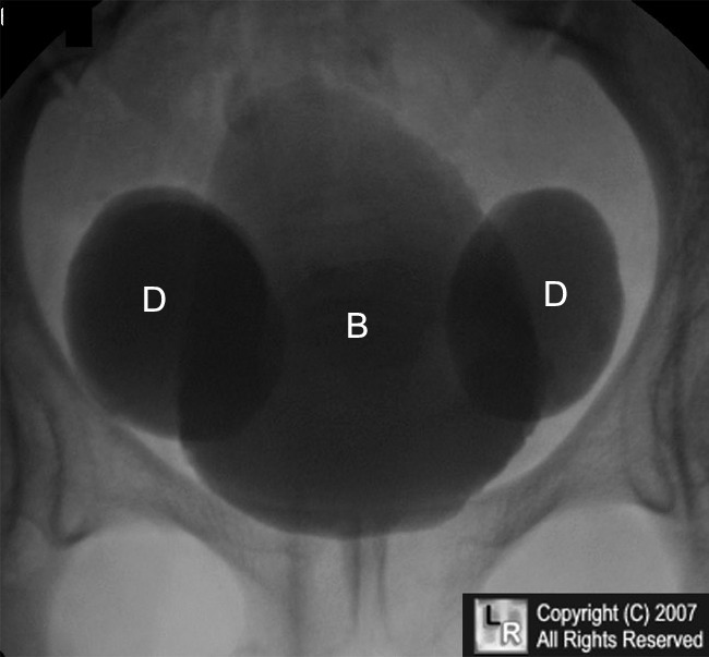
Large Bladder Diverticula. A view from a voiding cystourethrogram demonstrates two large bladder diverticula (D) flanking the bladder lumen (B). These diverticula are in the region of the insertion of the ureters. They are frequently associated with reflux (Hutch diverticula) but no reflux was demonstrated in this patient.
For additional information about this disease, click on this icon if seen above.
For this same photo without arrows, click here
“Bladder Anomalies” eMedicine Bartley G Cilento, MD
|
|
|
