|
|
Adrenal Calcifications
General considerations
- Hemorrhage is the most common cause
- Generally, patients with non-traumatic adrenal hemorrhage are critically ill and have a coagulopathy
- Adrenal glands are proportionately larger in neonates than adults and prone to complications from hypoxia or hypotension
- Unilateral 80% of time and involves right side 85% of time
- Unilateral adrenal hemorrhage rarely produces clinical findings
- In adults, it is frequently found incidentally in trauma
- Most (90%) of adrenal tissue must be destroyed for adrenal insufficiency
- Adrenal hemorrhage is prognostically important because it may signify severe stress or unsuspected coagulopathy
Adrenal Calcifications -- Causes |
Hemorrhage |
TB |
Waterhouse-Friderichsen Syndrome |
Neuroblastoma |
Pheochromocytoma |
Addison Disease |
Wolman Disease |
Adenoma |
Carcinoma |
Causes
- Hemorrhage
- More common than in adults
- May be associated with
- Birth trauma
- Hypoxia
- Septicemia
- Syphilis
- Adult
- Septicemia
- Waterhouse-Friderichsen Syndrome
- Septicemia, as from meningococcemia
- Hemorrhagic necrosis of adrenals from
- Septic emboli, or
- Disseminated intravascular coagulopathy
- Blunt abdominal trauma
- Adrenal may be compressed between spine and liver
- May undergo acute increase in adrenal venous pressure
- Shearing of small vessels may occur with deceleration
- Systemic coagulopathy
- Hemorrhage typically resolves in 4-6 weeks
- Addison Disease
- Primary adrenal insufficiency
- Affects adrenal cortex
- Adrenals are small
- Calcification can occur in 25% over a chronic course
- Infection
- Calcified granulomas in adrenal
- Histoplasmosis
- Calcified granulomas in adrenal
- Neoplasm
- Neuroblastoma
- Most common solid abdominal mass in infancy
- Almost always unilateral (90%)
- Stippled or coarse calcification is common (up to 70%)
- Pheochromocytoma
- Rare tumor of adrenal medulla
- May be bilateral (10%)
- About 10% calcify
- Adrenal cortical adenoma
- Unilateral
- Low attenuation (<10HU) on non-enhanced CT
- Calcification is rare
- Myelolipoma
- Unilateral
- Large, fat-containing adrenal
- About 20% calcify
- Wolman Disease
- Rare, autosomal-recessive metabolic disease leading to death in infancy
- Leads to abnormal collections of cholesterol esters and triglycerides in “foam cells”
- Diffuse punctate calcifications in adrenals which may be enlarged but maintain adreniform shape
- Hepatosplenomegaly
Imaging Findings
- CT is the study of choice in adults
- Adrenal hemorrhage and hemorrhagic adrenal rumors may appear similar
- Follow-up in 4 weeks to resolution for hemorrhage; tumors will not resolve
- Acute hemorrhage is hyperdense
- Associated with inflammatory stranding
- Adrenal is usually enlarged
- Bronchogenic carcinoma metastatic to the adrenals is the most common cause of an enlarged and hemorrhagic enhancing adrenal malignancy
- Ultrasound (US) is the modality of choice in neonates
- Sensitive to enlargement
- Glands may be large and hyperechoic early and hyperechoic centrally later
- MRI may be useful is evaluating the stage of hemorrhage
- On conventional radiography, adrenal calcifications will usually be bilateral and amorphous
- Located in the region of the adrenals, adjacent to the spine
- Calcification may be seen within weeks of the hemorrhage
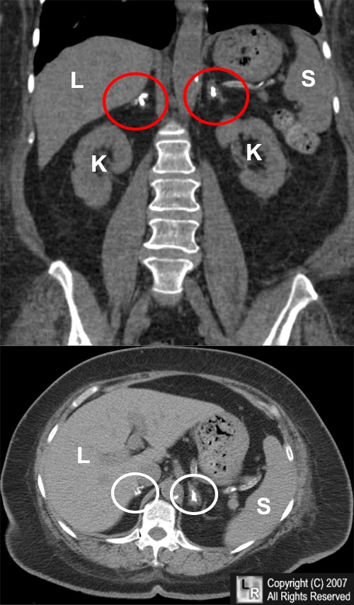
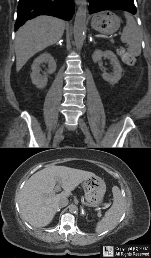
Bilateral adrenal calcifications. A coronal reformatted (above) and axial CT scan (below) of the upper abdomen show bilateral calcifications (red and white circles) in both adrenal glands. The adrenal glands are not enlarged. These calcifications were found incidentally and are most likely due to previous adrenal hemorrhage. The liver (L), spleen (S) and kidneys (K) are labeled.
For additional information about this disease, click on this icon if seen above.
For this same photo without the arrows, click here
|
|
|
