|
|
Osteitis Condensans Ilii
- General Considerations
- Benign sclerosis of the iliac side of the sacroiliac joints
- Bilaterally symmetric
- Radiologic diagnosis discovered serendipitously
- Usually found in young females who have had several children but also may be seen in males
- Related to remodeling of bone following stress across the sacroiliac joint
- Other theories of etiology are that it is related to a urinary tract infection, or that it
- May be related to a form of inflammatory arthritis
Clinical findings
- Rarely symptomatic but may be mildly painful
- May have morning stiffness and polyarthralgia
Imaging findings
- Sclerosis is frequently triangular in shape with the base pointing inferiorly
- Not to be confused with a sacroiliitis
- Sacroiliac joint is intact in osteitis condensans ilii
- Sclerosis involves only iliac side of joint in osteitis condensans ilii
- Outer margin of sclerosis is usually well-defined
- If in doubt, oblique views of the SI joints will show the joint to be intact and the sclerosis on only iliac side
- May spontaneously resolve

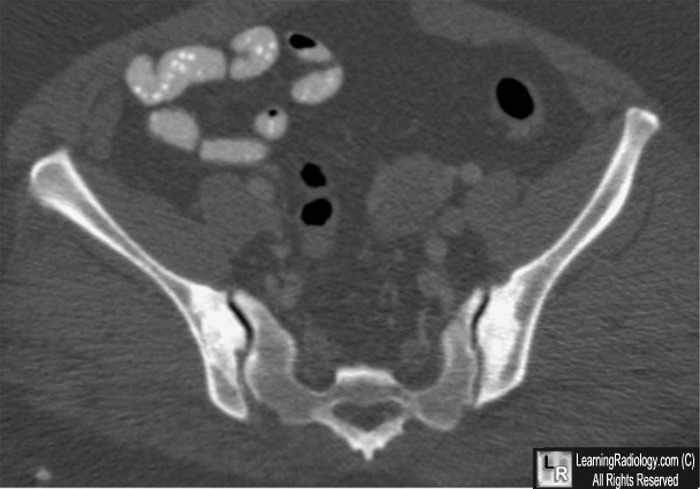
Osteitis Condensans Ilii. This is a single axial image from a CT scan of the pelvis demonstrating
sclerosis (blue arrows) on only the iliac side of the sacroiliac joints. The joints themselves are intact.
The sacral side of the joint is normal.
For this same photo without the arrows, click here
For more information, click on the link if you see this icon 
Osteitis condensans ilii WC Peh, MD Imaging Consultation American Journal of Orthopedics
|
|
|
