|
|
Mitral Annulus and Leaflet Calcification
- Mitral annulus frequently calcifies over the age of 60
- One of the most common cardiac calcifications
- Has been considered to be a degenerative process
- Calcification is actually subvalvular in location
- Associations
- Aortic stenosis
- Hypertrophic cardiomyopathy
- Chronic renal failure, especially of those on dialysis
- Bacterial endocarditis
- Systemic hypertension
- Diabetes mellitus
- Hypercholesterolemia
- It may be associated with significant atherosclerosis
- In one study, it was found to be associated with twice the risk of stroke, independent of other risk factors
- In another study, it was associated with an increased prevalence of coronary artery disease in patients <65
- Calcific deposits can lead to cardiac conduction disturbances
- Usually occurs over the age of 40, more often after 65-70
- More common in women
- Not clinically significant unless massive
- May lead to mitral insufficiency
- Shaped like a U, J or reverse C
- Appears as a band of increased density
- Echocardiography and CT are most sensitive means of imaging it
- Mitral leaflet calcifications
- Formerly calcification of the mitral valve itself was most commonly due to rheumatic fever
- Rheumatic fever produces valve abnormalities leading to stenosis and regurgitation, including:
- Calcification and thickening of leaflets and chordae tendineae
- Fusion of commissures
- Can also occur as a degenerative process
- Prevalence increases with age
- Found in 75% over age 80 on CT
- Not associated with mitral stenosis
- Whereas aortic valve calcifications are invariably associated with aortic stenosis
- When rheumatic in origin, mitral valve calcification is usually associated with mitral stenosis
- Imaging findings
- Heavier calcific deposits in men than women
- Not usually apparent on conventional radiography
- Calcium usually deposited in clumps on valve leaflets
- On a lateral chest radiograph, a line drawn connecting the carina and the anterior costophrenic sulcus will usually mark the location of the aortic valve above and the mitral valve below
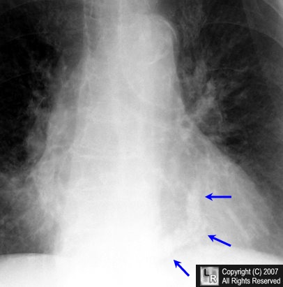
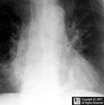
Calcification of the Mitral Annulus . The blue arrows point to dense, amorphous calcification
arranged in a
curvilinear path that corresponds to the location of the annulus of the mitral valve.
For the same photo without the arrows, click here
|
|
|
