|
|
Chopart Fracture-Dislocation
- The foot is generally divided into the
- Hindfoot
- Midfoot
- Cuboid
- Navicular
- Three cuneiforms
- Forefoot
- The articulation between the hindfoot and the midfoot (midtarsal joint) is frequently referred to as Chopart’s joint
- Named after surgeon who performed amputations at the calcaneocuboid, talonavicular joint
- Named after French surgeon Francois Chopart (1743–1795) who performed amputations of the foot at this level
- This type of amputation renders the ankle joint unstable as almost all of the points of insertion of the ankle tendons have been remove
- The articulation between the midfoot and the forefoot is referred to as the Lisfranc joint
- All dislocations of the foot are relatively uncommon with the Lisfranc fracture-dislocation being the most common
- Most are due to falls from a height or motor vehicle accidents
- Males are more likely to have foot dislocations than females
- Chopart Fracture
- Chopart fracture-dislocation involves the midtarsal joints (talonavicular and calcaneocuboid joints)
- Typically caused by falls from a height, motor vehicle accidents and severe twisting injuries such as can occur in basketball players who land on a plantar-flexed and inverted foot
- Usually result from severe trauma
- Most commonly, there is medial displacement of the distal fragments (80%)
- The foot is displaced inward and upward
- But displacement in other directions can occur
- Eversion injuries result in lateral dislocations
- There are frequently associated fractures of the calcaneus, cuboid and navicular
- A small percentage are open
- The talus remains in the ankle mortise
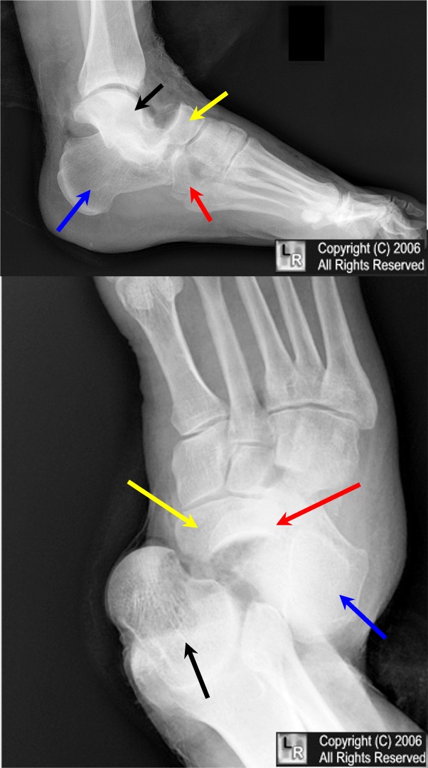
Chopart's fracture dislocation. Black arrow points to talus which is dislocated from navicular (yellow
arrow) at talonavicular joint. Calcaneus (blue arrow) is dislocated from the cuboid (red arrow), which is also fractured. The dislocation is at the calcaneocuboid joint.
This is an uncommon dislocation.
The forefoot is usually displaced medially rather than laterally as in this case.
For the same photo without the arrows, click here.
- Prompt reduction and early range of motion generally result in favorable outcome
- High impact injuries with greater soft tissue compromise and associated fractures worsen prognosis
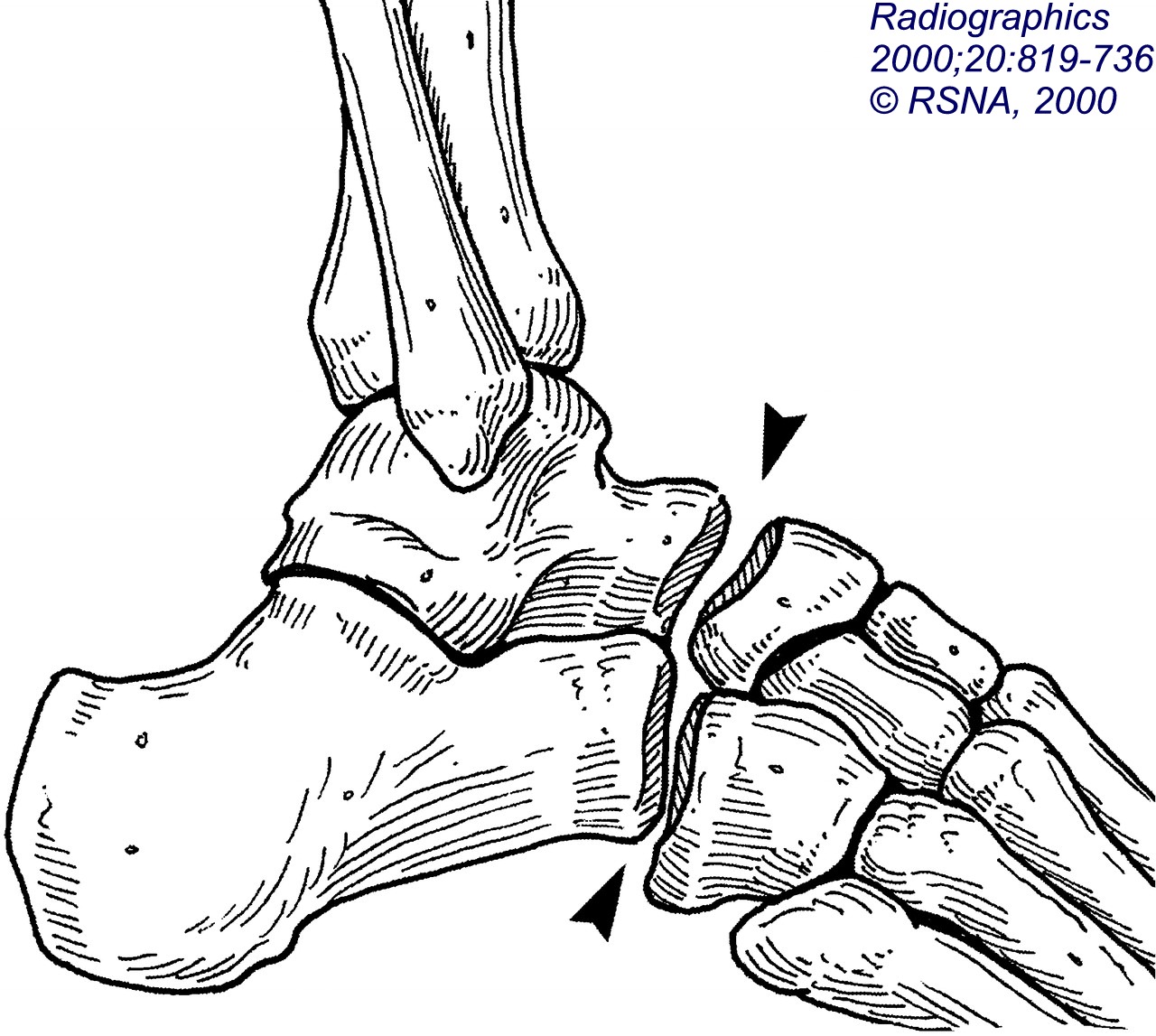 . .
Diagram of Chopart's fracture-dislocation from Radiographics
Radiographics. 2000;20:819-736. © RSNA, 2000 from: Radiologic History Exhibit 1: Musculoskeletal Eponyms: Who Are Those Guys? By Tim B. Hunter, MD, Leonard F. Peltier, MD, PhD and Pamela J. Lund, MD
American Journal of Roentgenology: 2004; 183:615-622 © American Roentgen Ray Society Ankle and Foot Injuries: Analysis of MDCT Findings by Ville V. Haapamaki, Martti J. Kiuru, and Seppo K. Koskinen
|
|
|
