|
|
Calcification of the Left Atrial Wall
General considerations
- Calcification of left atrium relatively common finding in patients with long-lasting rheumatic valve disease
- Massive calcification of the left atrial walls (porcelain atrium) is rare condition with implications for mitral valve surgery (see below)
- Massive calcification of the left atrium predominantly affects women (3/4 of cases)
- Almost always associated with rheumatic mitral stenosis
- Most patients have experienced long-term symptoms (more than 15 years)
- Most patients have previously undergone mitral valve operations
Almost all have atrial fibrillation
- With an average duration of 10 years
Location
- Usually spares the interatrial septum
Patterns of calcification
- Type A
- Calcification in the left atrial appendage only
- Underlying lesion is most commonly mitral stenosis
- Almost always associated with thrombus in the appendage
- Type B
- Free wall of the left atrium and mitral valve are calcified
- Indicates advanced mitral stenosis
- Type C
- Small area of calcification is confined to the posterior wall of the LA
- Results from a jet lesion because of mitral regurgitation
- Called a McCollum patch
Treatment
- Surgical technique during interventions for valvular substitution are difficult with calcification of the wall
- Dislodgement of thrombus from the left atrium during surgery can result in cerebral embolism and uncontrollable hemorrhage if the left atrium is entered through the calcified region
- This is because of wall rigidity
- Endarterectomy with mitral valve replacement is the currently accepted corrective procedure
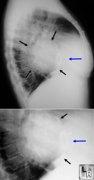
Calcification
of left
atrial
wall in
long-standing
mitral
stenosis. Upper
photo:
The left
atrium
(black
arrows)
is
located
in the
center
of the
heart
posteriorly.
The
anterior
wall of
the left
atrium
is
calcified
in this
photo
(see
inset in
lower
photo),
a
finding
usually
found in
patients
with
chronic
mitral
stenosis
who have
already
had
atrial
fibrillation
and a
mitral
valve
replacement.
There is
a
prosthetic
mitral
valve
present
(blue
arrows).
The
patient
had
mitral
stenosis
for 23
years.
Coconut Atrium: Transmural Calcification of the Entire Left Atrium. Carlos Del Campo, MD, Paul Weinstein, MD, Constantine Kunnelis, MD, Peter DiStefano, RDCS, RVT,* and Gloria M. Ebers, RT, CVT* Texas Heart Institute J. 2000; 27(1): 49–51. Copyright 2000 by the Texas Heart® Institute, Houston
eMedicine Cardiac Calcifications. Sohail G Contractor, MD with Pierre D Maldjian, MD, Mysore Seetharaman, MD, Hani H Abu-Judeh, MD, Farid Thanawala, MD
|
|
|
