|
|
Lipoma of the Cecum
- Uncommon tumors, but second in prevalence to adenomas for colonic tumors
- Tend to occur more frequently in older females
- Usually asymptomatic
- When symptomatic, can produce:
- Pain
- Diarrhea
- Rectal bleeding-if surface ulcerates
- Constipation
- Almost all are submucosal
- Most are located on the right side (40%), but about 20% are in the sigmoid
- In the small bowel, lipomas are more common proximally (duodenum)
- Imaging findings
- Usually less than 4 cm in size
- Smooth, sharply defined hemispheric mass
- Typically produces either right-angle or slightly obtuse angle as the lesion meets lumen of bowel
- Rarely pedunculated
- Squeeze-sign = deformity due to softness and compressibility of these lesions
- Contour may be altered by peristalsis
- Ulceration is rare
- CT may demonstrate fatty nature of lesion, especially if they are large enough for accurate density measurements
- May intussuscept
- Do not undergo malignant transformation
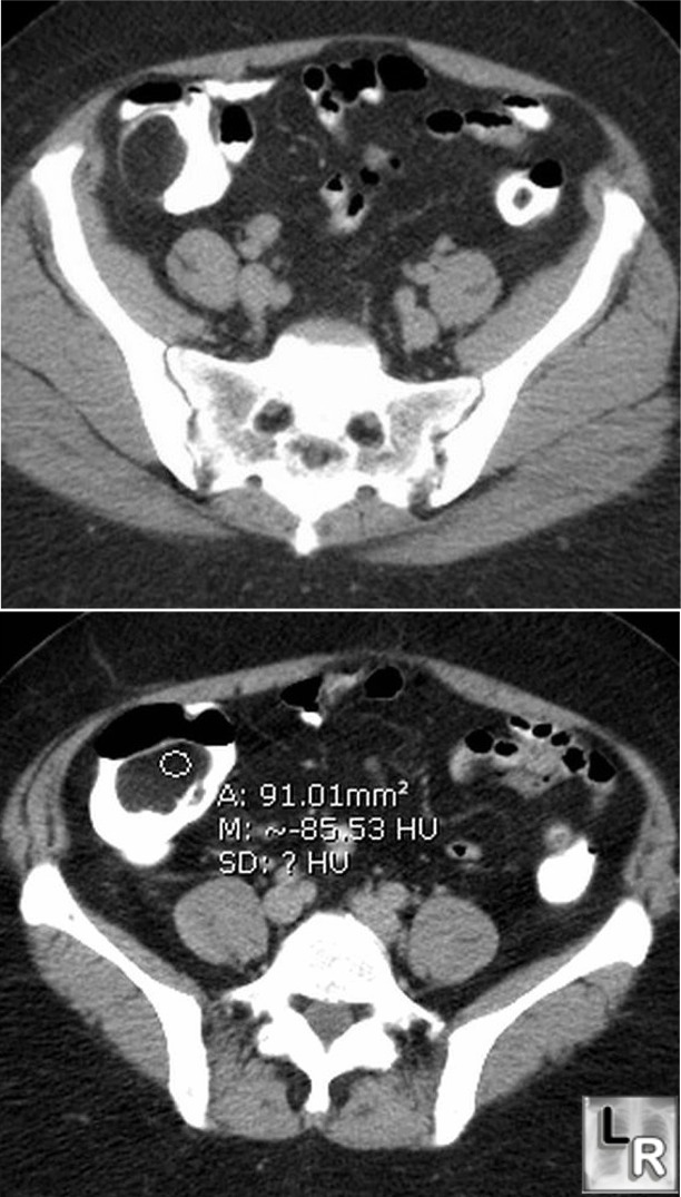
Cecal Lipoma. Axial CT image of right lower
quadrant shows a large, lobulated
filling defect in the cecum
with well-circumscribed margins.
The lower image demonstrates a
negative Hounsfield values (-85HU)
consistent with fat. The lesion
represents a lipoma of the cecum.
|
|
|
