|
|
Acromioclavicular Separation
AC Separation
- Normal anatomy of acromioclavicular (AC) joint
- Synovial joint
- Acromioclavicular ligament
- Coracoclavicular ligaments
- Medial conoid and lateral trapezoid
- Denser, thicker, stronger
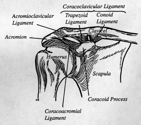
From Harris and Harris, Radiology of Emergency Medicine
- Normal measurements
- AC joint space is usually <5mm
- Right and left differ by no more than 2-3
mm
- Coracoclavicular distance usually <11-13 mm
- Right and left should differ by < 5 mm
- 50% difference in size between the two
shoulders is considered significant
- Inferior plane of the distal clavicle should
be on same plane as inferior border of acromion
- Developmental variations reported as high
as 19%
- Fall on shoulder is frequent mechanism of
injury
- Point tenderness, limitation of motion
- Abnormal widening of the AC joint due to
disruption of the AC ligament
- CC separation is the more important soft
tissue injury
- Extent of CC separation has direct effect on
degree of AC separation
- Classification
Type |
Anatomy |
Radiographic findings |
Prognosis |
I
Sprain |
Stretching
of AC ligament
AC joint is
stable
CC ligament
intact
|
Only seen
on stress views of injured and uninjured shoulders=widening of
AC joint |
No
instability |
II
Subluxation |
Partial or
complete rupture of AC ligament
Partial,
but not complete, disruption of CC ligament
|
Widening of
AC joint but a normal coracoclavicular distance
Stress
films may still be required to demonstrate widening of both AC
joint and CC space
|
May require
arthroplasty |
III |
Disruption
of both AC and CC ligaments |
Widening of
both the AC and CC spaces on routine erect film
|
Internal fixation |
IV
Posterior |
AC and CC
ligaments disrupted but coracoacromial ligament remains intact
|
Distal end
of clavicle lies inferior and posterior to acromion seen best on
axillary view |
|
V
Inferior |
AC and CC
ligaments disrupted
Coracoacromial ligament remains intact Sternoclavicular
separation occurs as well
|
Marked
widening of both the AC and CC space
Sternoclavicular dislocation |
|
VI |
Distal end
of clavicle displaced inferiorly and lodges in biceps and
coracobrachialis muscles
|
Distal end
of clavicle comes to lie inferior to acromion |
|
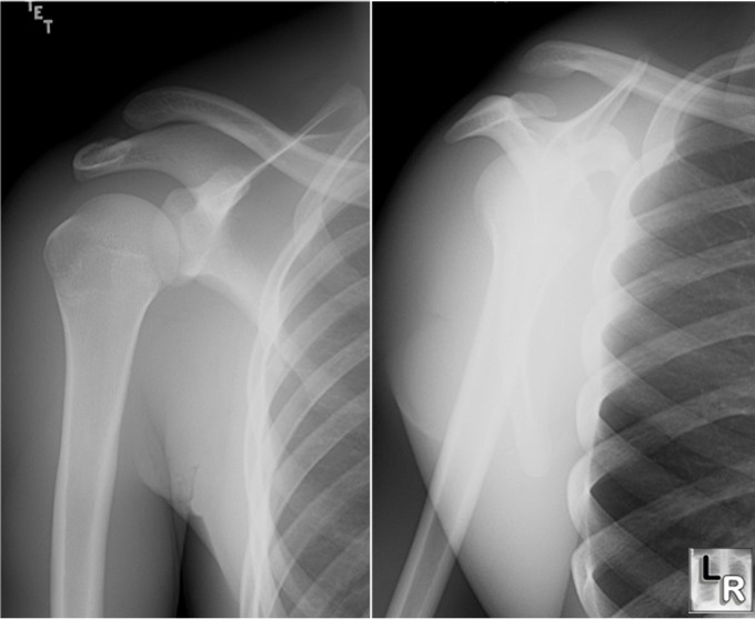
Two views of right shoulder show elevation of the clavicle
and separation of the AC joint in a Type III AC joint separation
- Fracture of distal end of clavicle is
frequently associated with CC tears with or without separation of AC
ligament
- Separation may heal with soft tissue
calcification or ossification
Harris, J and Harris, W: Radiology of
Emergency Medicine, 4th ed,
2000.
Manaster,
B., Disler, D.,May,
D.: The Requisites: Musculoskeletal Imaging, 2002
|
|
|
