|
|
Osteochondritis Dissecans
-
Sub-articular,
post-traumatic necrosis
-
Occurs only
on convex surfaces of bone
-
Most patients
are athletic
-
Direct blow
is more common cause than a rotational injury
-
Most common
cause of an intra-articular loose body
-
In adults,
loose body contains larger fragment of cartilage than bone
-
Possible
outcomes
-
Death of bony,
but not cartilaginous, portion of loose body
-
Complete resorption
of loose body
-
Reincorporation
or regrowth
-
Cause of a
“locking knee”
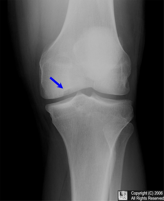
Osteochondritis dissecans. Blue arrow points to crescentric lucency in the convex
surface of the medial condyle of the knee.
For the same photo without the arrows, click here.
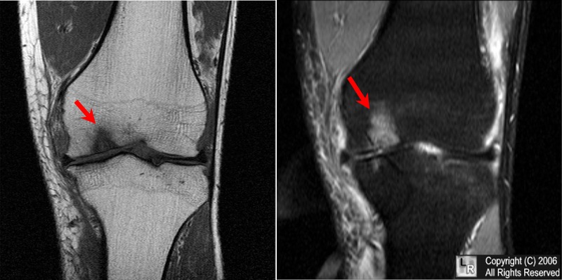
Osteochondritis dissecans. Red arrows point to osteochondral defect and bone edema on T1 and stir
MRI images of the knee in same patient as above.
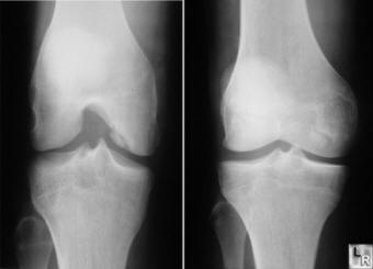
Osteochondritis dissecans of medial femoral
condyle-ovoid fragment
of bone is separated from surface of condyle but does not yet lie
freely within the joint.
|
|
|
