|
|
Pulmonary Arterial Hypertension
- Sustained pulmonary arterial pressure
- In systole >30 mm Hg
- In diastole >15 mm Hg
- Mean pressure >20 mm Hg
- Pathogenesis:
- Primary PAH (rare) = unknown cause
- Diagnosis of exclusion
- Clinically unexplained progressive pulmonary arterial hypertension without evidence for thromboembolic disease and pulmonary venoocclusive disease
- Clinical
- Age
- 3rd decade; females > males
- Dyspnea on exertion
- Syncope
- Easy fatigability
- Hyperventilation
- Chest pain
- Imaging findings
- Main pulmonary artery usually prominent
- Right and left pulmonary arteries large and taper rapidly
- Peripheral pulmonary arteries are narrow and inconspicuous
- Diffuse oligemia of the lungs
- No overinflation
- Nuclear medicine
- Diffuse patchy defects on perfusion scans (low probability)
- On CT, main pulmonary artery diameter should be less than 30mm
- Secondary PAH (more common)
- Primary pleuropulmonic disease
- Parenchymal pulmonary disease
- COPD
- Emphysema
- Chronic bronchitis
- Asthma
- Bronchiectasis
- Granulomatous disease
- Cystic fibrosis
- End-stage fibrotic lung
- S/P lung resection
- Idiopathic hemosiderosis
- Alveolar proteinosis
- Alveolar hypoventilation = hypoxic pulmonary arterial hyperperfusion
- Chronic high altitude
- Sleep apnea
- Hypoventilation due to neuromuscular disease or obesity
- Pleural disease and chest deformity
- Fibrothorax
- Thoracoplasty
- Kyphoscoliosis
- Primary vascular disease
- Congenital heart disease
- Increased flow: large L-R shunt
- Decreased flow: Tetralogy of Fallot
- Capillary obliteration
- Chronic pulmonary thromboembolism
- Persistent fetal circulation
- Arteritides (eg, Takayasu)
- Venous obliteration
- Pulmonary venoocclusive disease
- Pulmonary venous hypertension
- Prognosis
- Majority of patients have a median survival of 2-3 years
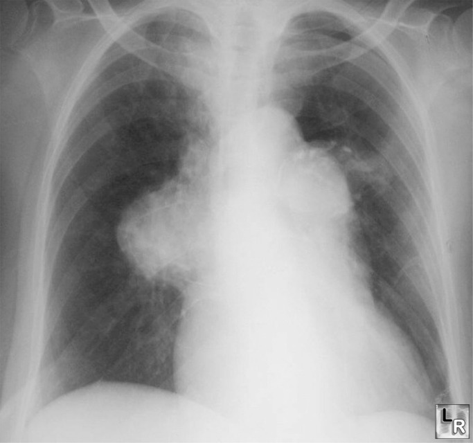
Pulmonary Arterial Hypertension. Frontal radiograph of the chest shows an enlarged main pulmonary
artery
and a markedly enlarged right and left pulmonary arteries. The
peripheral vasculature is
normal.
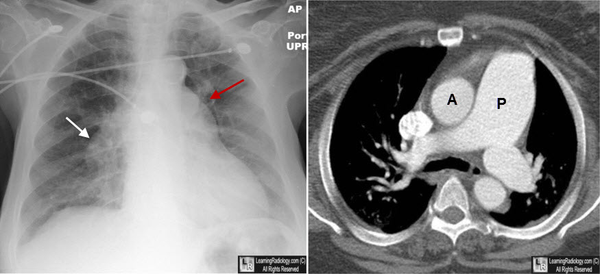
Pulmonary Arterial Hypertension. Frontal radiograph of the chest shows an enlarged main pulmonary
artery (red arrow) and a markedly enlarged right pulmonary artery (white arrow) with rapid attenuation in the size of the vessels in the lung periphery. On the contrast-enhanced CT, the main pulmonary artery (P) is much larger than the aorta (A). Normally, they should roughly be about the same size. The main pulmonary artery should not measure more than 3 cm in diameter on CT.
|
|
|
