|
|
Penetrating Aortic Ulcer
Submitted by Anthony Chang, MD
- Ulceration of an atherosclerotic plaque which penetrates into the internal elastic lamina
- Hematoma then forms within the media of the aortic wall
- Occurs in the elderly who usually have a history of severe atherosclerosis, hypertension, and hyperlipidemia
- Similar presentation to those with a descending thoracic aortic dissection i.e. acute chest or back pain
- Plaque ulceration usually in the middle to distal third of the descending aorta
- Intramural hematoma accompanies the penetrating ulcer 80% of the time
- Associated with abdominal aortic aneurysm
- Disease progresses from intimal plaque ulceration to media hematoma formation to adventitial saccular pseudoaneurysm formation and finally rupture if there is transmural penetration
- Speculated as the cause of descending or thrombosed type dissections with all three
Radiographic findings
- Focal contrast collection projecting beyond the aortic lumen on CT
- Intramural hematoma is indistinguishable from intraluminal thrombus
- Intimal flap is uncommon
- Intramural wall thickening or thrombus is frequently found
- On angiography, there is aortic wall thickening and the ulcerated plaque seen
- On MRI
- High signal intensity on both T1 and T2 with subacute hematoma
- Can be demonstrated by computed tomography, magnetic resonance, angiography and trans-esophageal echocardiography
- Differential diagnosis:
- Aortic dissection (has an intimal flap)
- Atheroma – has a low signal on both T1 and T2
Treatment
- Surgical cases are those demonstrating hematoma expansion, impending rupture, inability to control blood pressure
- Patients routinely have co-morbid conditions that make them poor surgical candidates and are treated with transluminal placement of endovascular stent grafts
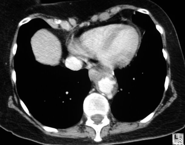
Penetrating Aortic Ulcer. Enhanced CT scan through the lower thoracic aorta demonstrates
a focal outpouching of contrast posteriorly representing a
penetrating aortic ulcer
|
|
|
